Trombosis venosa profunda
Revisado por Dr Colin Tidy, MRCGPÚltima actualización por Dr Toni Hazell, FRCGPÚltima actualización 15 Sept 2023
Cumple con las directrices editoriales
- DescargarDescargar
- Compartir
- Language
- Discusión
- Versión en audio
- Agregar a fuentes preferidas en Google
En esta serie:Cómo prevenir la TVP al viajar
Una trombosis venosa profunda (TVP) es un coágulo de sangre en una vena. Los coágulos de sangre en las venas ocurren con mayor frecuencia en las piernas, pero pueden ocurrir en otras partes del cuerpo, incluidos los brazos. Este folleto trata sobre los coágulos de sangre en las venas de las piernas.
La causa más común de que se desarrolle un coágulo de sangre en una vena es la inmovilidad. En algunos casos, puede ocurrir una complicación donde parte del coágulo de sangre se desprende y viaja al pulmón (embolia pulmonar). Esto generalmente se previene si se le administra tratamiento anticoagulante.
De un vistazo
Una trombosis venosa profunda (TVP) es un coágulo de sangre que se desarrolla en una de sus venas profundas, generalmente en la pierna.
Los síntomas comunes incluyen dolor, sensibilidad e hinchazón de la pantorrilla, que también puede volverse caliente y roja.
La causa más común de una TVP es la inmovilidad, especialmente durante o después de una cirugía.
Una TVP puede llevar a una embolia pulmonar (EP) si parte del coágulo viaja a los pulmones.
El tratamiento incluye medicamentos anticoagulantes, medias de compresión, descansar con las piernas elevadas y mantenerse activo.
El movimiento regular, especialmente durante viajes largos o después de una cirugía, ayuda a prevenir los trombos venosos profundos (TVP).
¿Qué es una trombosis venosa profunda?
A deep vein thrombosis (DVT) is a blood clot that develops in one of your deep veins. It usually occurs in your leg and makes that leg become very painful and swollen. The clot may travel up to your lungs to cause a embolia pulmonar.
Diagrama de la pierna que muestra las venas y una Trombosis Venosa Profunda
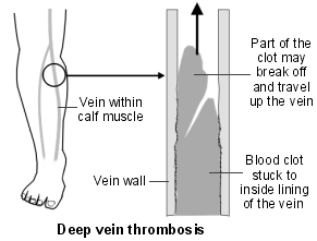
Deep leg veins are the larger veins that go through the muscles of the calf and thighs. They are not the veins that you can see just below the skin; neither are they the same as las venas varicosas. Cuando tienes una TVP, el flujo sanguíneo en la vena está parcial o completamente bloqueado por el coágulo de sangre.
Una vena de la pantorrilla es el sitio común para una TVP. Una vena del muslo se ve afectada con menos frecuencia. Raramente, otras venas profundas en el cuerpo pueden ser bloqueadas por coágulos de sangre.
Una TVP es parte de un grupo de problemas conocidos en conjunto como tromboembolismo venoso.
Causas de una trombosis venosa profunda
La sangre normalmente fluye rápidamente a través de las venas y no suele solidificarse (coagularse). El flujo sanguíneo en las venas de las piernas es facilitado por los movimientos de las piernas, ya que la acción muscular comprime las venas. A veces, una TVP ocurre sin razón aparente. Sin embargo, lo siguiente aumenta su riesgo de tener una TVP:
Inmovilidad which causes blood flow in the veins to be slow. Slow-flowing blood is more likely to clot than normal-flowing blood and those who are admitted to hospital should be assessed for their DVT risk soon after admission, and preventative measures put in place if needed.
Una operación quirúrgica donde estás dormido (bajo anestesia general) is the most common cause of a DVT. Your legs are still when you are under anaesthetic, because the muscles in your body are temporarily paralysed. Blood flow in the leg veins can become very slow, making a clot more likely to occur. Certain types of surgery (particularly operations on the pelvis or legs) increase the risk of DVT even more.
Cualquier enfermedad o lesión que cause inmovilidad increases the risk of DVT. This includes having a leg in a hard plaster cast after a fracture. People who are admitted to intensive care units are at an increased risk of DVT. This is due to a number of reasons but partly because they are very ill and also because they are immobile (they may even be kept asleep by anaesthetic medications). Those being discharged from hospital with a problem that affects movement of the legs should also have a risk assessment and treatment if appropriate.
Largos viajes en avión, tren o autobús/coche may cause a slightly increased risk. This is because you are mostly sitting for long periods of time and not moving around very much.
Daño al revestimiento interno de la vena increases the risk of a blood clot forming. For example, a DVT may damage the lining of the vein. So, if you have a DVT then you have an increased risk of having another one in the future. Some conditions, such as inflammation of the vein wall (vasculitis) and some medicines (for example, some chemotherapy medicines), can damage the vein and increase the risk of having a DVT. Damage to the vein can also happen with injury to the vein caused by a needle. This might occur after treatment with a drip in hospital (where a tube is inserted into a vein to get fluids into you). Drug users who inject drugs, such as heroin, can also damage their veins, making DVTs more common. This is especially the case if they inject illegal drugs into their leg or groin.
Condiciones que hacen que la sangre se coagule más fácilmente de lo normal (thrombophilia) can increase the risk. Some conditions can cause the blood to clot more easily than usual. Examples include nephrotic syndrome and antiphospholipid syndrome. See the separate leaflets called Síndrome Nefrótico, Síndrome Antifosfolípido y Trombofilia for more details. Some rare inherited conditions can also cause the blood to clot more easily than normal. An example of an inherited blood disorder which can cause DVTs is factor V Leiden mutation. You are more at risk of a DVT if you have a family history of DVTs - that is, a close relative who has had one.
Píldoras anticonceptivas, parches y anillos y terapia de reemplazo hormonal (TRH) which contain oestrogen can cause the blood to clot slightly more easily. Women taking the combined oral contraceptive (COC) pill or tablet HRT have a small increased risk of DVT. HRT which is transdermal (given through the skin as a patch, gel or spray) does not carry an increased risk of DVT, and nor does vaginal oestrogen given as part of HRT.
Personas con cáncer o insuficiencia cardíaca have an increased risk. Sometimes a DVT happens in a person who has not yet been diagnosed with cancer. Investigations looking for the cause of a DVT may show cancer to be the underlying cause.
Personas mayores (más de 60 años de edad) are more likely to have a DVT, particularly if they have poor mobility or have a serious illness such as cancer.
Embarazo increases the risk. About 1 in 1,000 pregnant women have a DVT while they are pregnant, or within about six months after they give birth.
Obesidad and being overweight also increases the risk. If your índice de masa corporal (IMC) is more than 25 kg/m2, tienes más riesgo de TVP.
Falta de líquidos en el cuerpo (deshidratación) can make a DVT more likely to happen. Effectively, the blood becomes stickier and more liable to clot.
¿Qué tan común es una trombosis venosa profunda?
Se estima que aproximadamente 1 de cada 1,000 personas tiene una TVP cada año en el Reino Unido.
Síntomas de trombosis venosa profunda
Una TVP se desarrolla más comúnmente en una vena profunda debajo de la rodilla en la pantorrilla. Los síntomas típicos de la TVP incluyen:
Dolor y sensibilidad en la pantorrilla.
Hinchazón de la pantorrilla.
Cambios de color y temperatura de la pantorrilla. La sangre que normalmente pasaría por la vena bloqueada se desvía hacia las venas externas. La pantorrilla puede entonces volverse cálida y roja.
A veces no hay síntomas y una TVP solo se diagnostica si ocurre una complicación, como una embolia pulmonar (EP) - ver abajo.
Diagnóstico de una trombosis venosa profunda
Sometimes it is difficult for a doctor to be sure of the diagnosis from just your symptoms, as there are other causes of a painful and swollen calf. Examples of conditions that can cause síntomas similares symptoms are distensiones musculares o infecciones de la piel (celulitis).
Su médico podría calcular algo llamado puntuación de Wells para determinar la probabilidad de que tenga una TVP. Esto implica observar sus síntomas y factores de riesgo para una TVP. El médico también considerará si hay algún síntoma o signo que sugiera que el coágulo ha viajado a sus pulmones y ha causado una EP.
Si se sospecha de una TVP, normalmente se le aconsejará realizar pruebas de manera urgente para confirmar o descartar el diagnóstico. Dos pruebas comúnmente utilizadas son:
Prueba de sangre de dímero D
Esto detecta fragmentos de productos de descomposición de un coágulo de sangre. Cuanto más alto sea el nivel, más probable es que tenga un coágulo de sangre en una vena. Desafortunadamente, la prueba puede ser positiva en varias otras situaciones, como si ha tenido una cirugía reciente o si está embarazada.
Por lo tanto, una prueba positiva no diagnostica una TVP. Sin embargo, la prueba puede indicar qué tan probable es que tenga un coágulo de sangre (el coágulo puede ser una TVP o una EP) y puede ayudar a decidir si se necesitan más pruebas.
Doppler Duplex
Este tipo especial de ecografía se utiliza para mostrar el flujo sanguíneo en las venas de las piernas y cualquier obstrucción al flujo sanguíneo. La ecografía es útil porque es una prueba fácil y no invasiva. Idealmente, esta prueba debería realizarse dentro de las cuatro horas; esto a menudo no es prácticamente posible, en cuyo caso se puede administrar una inyección de medicamento anticoagulante mientras espera la exploración.
Otras pruebas
A veces, estas pruebas no son concluyentes y se necesitan pruebas más detalladas. La venografía de contraste es otra prueba que se puede realizar. En esta prueba se inyecta un tinte en las venas de las piernas.
X-ray tests can then detect the dye which is shown not to be flowing if a vein is blocked by a clot. Tomografías computarizadas (TC) y escáneres de resonancia magnética (RM) are also very occasionally needed.
Complicaciones de una trombosis venosa profunda
Cuando se forma un coágulo de sangre en una vena de la pierna, generalmente permanece adherido a la pared de la vena. Los síntomas tienden a estabilizarse gradualmente. Sin embargo, hay dos posibles complicaciones principales de la TVP:
Un coágulo de sangre que se desprende y viaja al pulmón (embolia pulmonar).
Síntomas persistentes en la pantorrilla (síndrome postrombótico).
Embolia pulmonar (EP)
In a small number of people who have a DVT, a part of the blood clot breaks off. This travels in the bloodstream and is called an embolus. An embolus will travel in the bloodstream until it becomes stuck. An embolus that comes from a clot in a leg vein will be carried up the larger leg and body veins to the heart, through the large heart chambers, but will get stuck in a blood vessel going to a lung. This is called a embolia pulmonar.
Los DVT y PE se conocen colectivamente como tromboembolismo venoso
Un pequeño EP puede no causar ningún síntoma. Un EP de tamaño mediano puede causar problemas respiratorios y dolor en el pecho. Un EP grande puede causar colapso y muerte súbita. Se estima que si las personas con TVP no son tratadas, aproximadamente la mitad de ellas desarrollaría un EP lo suficientemente grande como para causar síntomas o la muerte.
Síndrome postrombótico
Without treatment, up to 5 in 10 people who have a DVT develop long-term symptoms in the calf. This is called síndrome postrombótico. Los síntomas ocurren porque el aumento del flujo y la presión de la sangre desviada hacia otras venas puede afectar los tejidos de la pantorrilla.
Los síntomas pueden variar de leves a graves e incluyen dolor en la pantorrilla, malestar, hinchazón y erupciones. En casos graves, puede desarrollarse una úlcera en la piel de la pantorrilla.
El síndrome postrombótico es más probable que ocurra si la TVP se presenta en una vena del muslo o se extiende hacia una vena del muslo desde una vena de la pantorrilla. También es más común en personas con sobrepeso u obesidad, en aquellas que son mayores y en aquellas que han tenido más de una TVP en la misma pierna.
Objetivos del tratamiento para una trombosis venosa profunda
Los objetivos del tratamiento son:
Para evitar que el coágulo se extienda por la vena y se haga más grande. Esto puede prevenir que un gran émbolo se desprenda y viaje a los pulmones (una EP).
Para reducir el riesgo de desarrollar síndrome postrombótico.
To reduce the risk of úlceras venosas in the leg in future. This can happen to people who have developed post-thrombotic syndrome.
Para reducir el riesgo de una futura TVP en el futuro.
Tratamiento de la trombosis venosa profunda
Anticoagulación - prevenir que el coágulo se haga más grande
La anticoagulación a menudo se llama adelgazamiento de la sangre. Los medicamentos que funcionan de esta manera se llaman anticoagulantes. Sin embargo, no adelgazan la sangre realmente. Alteran ciertos químicos en la sangre para evitar que los coágulos se formen tan fácilmente. Los anticoagulantes no disuelven el coágulo.
La anticoagulación evita que una TVP se haga más grande y previene la formación de nuevos coágulos. Los propios mecanismos de curación del cuerpo pueden entonces comenzar a trabajar para descomponer el coágulo.
Si tienes una TVP, generalmente necesitarás un medicamento anticoagulante durante al menos tres meses y luego deberías tener una revisión con un especialista. Hay varias opciones:
Warfarina has been the usual anticoagulant for many years. The aim is to get the dose of warfarin just right so the blood will not clot easily, but not too much, which may cause bleeding problems. You will need regular blood tests (called INRs) whilst you take warfarin. The INR (which stands for International Normalised Ratio) is a blood test that measures your blood clotting ability. You need the tests quite often at first but then less frequently once the correct dose is found. An INR of 2.5 is the aim if you have warfarin for a DVT, although anywhere in the range 2-3 is OK. If you have had recurrent DVTs, or have had a PE whilst on warfarin, you might need a higher INR (even 'thinner' blood). Recently, some alternatives to warfarin have become available, which do not need regular blood tests.
Rivaroxaban, apixaban, edoxabany dabigatran are newer anticoagulant medicines. You do not have to have regular blood tests to monitor your blood clotting. This is an advantage over warfarin. However, there is no antidote (as there is with warfarin) to stop you bleeding too easily. These newer tablets are not suitable for everybody.
NICE recomienda:
Apixaban o rivaroxaban para TVP o EP confirmada.
Si ni apixabán ni rivaroxabán son adecuados, entonces:
Inyecciones de heparina de bajo peso molecular (HBPM) durante al menos 5 días seguidas de dabigatrán o edoxabán, o
Inyecciones de LMWH con Warfarina durante al menos 5 días, seguidas de Warfarina sola.
Un émbolo grave es raro si comienzas el tratamiento anticoagulante temprano después de una TVP.
Si estás embarazada, se pueden usar inyecciones regulares de heparina en lugar de tabletas anticoagulantes. Esto se debe a que los medicamentos anticoagulantes pueden causar daño (defectos de nacimiento) al feto.
El tiempo que se le aconsejará tomar anticoagulación depende de varios factores. Si tiene una trombosis venosa profunda (TVP) durante el embarazo o después de una operación, entonces después del parto, o cuando esté en forma nuevamente, el riesgo aumentado se reduce considerablemente. Por lo tanto, la anticoagulación puede ser solo por unos pocos meses. Por otro lado, algunas personas continúan teniendo un riesgo aumentado de tener una TVP. En este caso, la anticoagulación puede ser a largo plazo.
Como guía, para una TVP que ocurre por debajo de la rodilla, necesitarás al menos seis semanas de tratamiento anticoagulante. Necesitarás al menos tres meses de tratamiento si la TVP está por encima de la rodilla. Algunas personas con mayor riesgo de otra TVP pueden necesitar más tiempo que esto. La duración del tiempo de anticoagulación varía de persona a persona. Tu médico o la clínica de anticoagulantes te aconsejarán cuánto tiempo durará tu tratamiento.
Medias de compresión
A algunas personas que desarrollan una TVP se les aconseja usar medias de compresión. Las medias de compresión también se conocen como medias de compresión o medias de prevención de tromboembolismo ('TEDs'). Estas se utilizan solo para tratar síntomas continuos; solían recomendarse para reducir el riesgo de desarrollar otra TVP o para reducir el riesgo de síndrome postrombótico, pero esto ya no se recomienda.
Las medias funcionan aplicando presión desde el exterior de las venas. Esto ayuda a exprimir la sangre en las venas hacia arriba por las piernas. Esto luego reduce la presión dentro de las venas. A su vez, esto previene la fuga de sangre hacia los tejidos circundantes.
Antes de recomendar medias de compresión, es posible que necesite hacerse una prueba para verificar que la circulación a través de las arterias de sus piernas sea normal. Esto generalmente lo realiza una enfermera, con una máquina portátil llamada Doppler. Esta mide la presión en sus arterias.
Las medias de compresión vienen en material ligero, medio o fuerte (clase 1, 2 o 3) y se usan por debajo de la rodilla. La mayoría de las personas pueden tolerar la clase 2. Si la clase 2 es demasiado incómoda o difícil, se prescribe la clase 1. Si las medias de clase 2 no funcionan, puede ser necesario probar la clase 3, aunque muchas personas encuentran estas difíciles de usar por cualquier período de tiempo.
Estos pueden ser recetados por su médico y obtenidos en la farmacia.
Consejos adicionales sobre medias de compresión
Están disponibles en diferentes colores, así que pide el que mejor te convenga. De esa manera, es más probable que los uses.
Deben quitarse a la hora de acostarse y ponerse a primera hora de la mañana. Es importante ponérselos antes de que las piernas comiencen a hincharse por la mañana.
Hay opciones de punta abierta o cerrada. Las medias de punta abierta pueden ser útiles si:
Tienes dolor en los dedos de los pies debido a la artritis o una infección.
Tienes pies grandes.
Quieres usar calcetines sobre tus medias de compresión.
Las prefieres.
Las medias deben ser reemplazadas cada 3-6 meses. Cada vez debes ser medido nuevamente, por si acaso es necesario cambiar la talla.
Siempre debes tener al menos dos pares prescritos para que un par pueda ser lavado y secado mientras el otro se usa.
No seque las medias de compresión en secadora, ya que esto puede dañar el elástico.
Pueden hacerse a medida si ninguna de las tallas estándar te queda bien. Esto aún se puede hacer con una receta.
Existen ayudas para aplicar medias de compresión si no puedes ponértelas. Puedes discutir esto con tu farmacéutico o enfermera.
Caminar regularmente pero elevar la pierna mientras descansas
A menos que su médico le aconseje lo contrario, debe caminar regularmente después de ser dado de alta del hospital. Se cree que caminar mejora la circulación en la pierna afectada y puede ayudar a reducir su riesgo de sufrir más trombosis venosa profunda (TVP).
Cuando estés descansando, tanto como sea posible, eleva tu pierna. Esto reduce la presión en las venas de la pantorrilla y ayuda a prevenir que la sangre y los fluidos se acumulen en las pantorrillas. Elevada significa que tu pie está más alto que tu cadera, por lo que la gravedad ayuda al flujo sanguíneo a regresar desde la pantorrilla. La forma más fácil de elevar tu pierna es recostarte en un sofá con la pierna sobre un cojín. Sentarse en el sofá o en una silla, con los pies en un reposapiés o puf, no es mantener los pies elevados, ya que los pies están muy por debajo de tus caderas en esta posición.
Eleva el pie de la cama unos centímetros si te resulta cómodo dormir así. Esto es para que tu pie y pantorrilla estén ligeramente más altos que tu cadera cuando estés dormido.
Otros tratamientos
A veces se pueden considerar otros tratamientos, por ejemplo:
Terapia trombolítica con medicamentos como estreptoquinasa o uroquinasa. Estos pueden ayudar a disolver un coágulo de sangre. El medicamento se administra directamente en la vena y, a veces, directamente al coágulo mediante un tubo (catéter). Los estudios informan que este tratamiento resulta en menos personas desarrollando síndrome postrombótico. Aún no se sabe si hace que las personas sean menos propensas a tener una EP, u otra TVP.
A veces se realiza una operación (llamada embolectomía) para eliminar un coágulo de sangre de la vena de la pierna o de la arteria pulmonar. Estas operaciones no son rutinarias y no está claro si son un tratamiento efectivo en la mayoría de los casos.
Ocasionalmente, se realiza una operación para colocar un filtro en la vena grande por encima de la vena bloqueada de la pierna. El objetivo es detener cualquier coágulo de sangre que pueda viajar hacia los pulmones. Esto puede considerarse si no se puede administrar anticoagulación (por diversas razones), o si la anticoagulación no logra prevenir que los coágulos se desprendan y viajen hacia las venas más grandes y hasta los pulmones.
Prevención de una primera aparición o recurrencia de trombosis venosa profunda
Una TVP a menudo es solo un evento aislado después de una operación mayor.
Sin embargo, algunas personas que desarrollan una TVP tienen un riesgo continuo de desarrollar otra TVP; por ejemplo, si tienes un problema de coagulación sanguínea o inmovilidad continua. Como se mencionó anteriormente, es posible que se te aconseje tomar medicamentos anticoagulantes a largo plazo. Tu médico te aconsejará sobre esto.
Otras cosas que pueden ayudar a prevenir una primera o recurrente TVP incluyen lo siguiente:
Si es posible, evita largos períodos de inmovilidad, como estar sentado en una silla durante muchas horas. Si puedes, levántate y camina de vez en cuando. Un paseo diario enérgico de 30-60 minutos es aún mejor si puedes hacerlo. El objetivo es evitar que la sangre se acumule y mejorar la circulación en las piernas. El ejercicio regular de los músculos de la pantorrilla también ayuda. Puedes hacer algunos ejercicios de pantorrilla incluso cuando estás sentado.
Las operaciones mayores son un riesgo para una TVP, especialmente las operaciones en la cadera, el abdomen inferior y la pierna. Hay una serie de métodos para ayudar a reducir este riesgo:
Para ayudar a prevenir una TVP, es posible que se le administre un anticoagulante, como una inyección de heparina, justo antes de una operación. Esto se llama profilaxis. La enoxaparina y la dalteparina son los tipos más comunes de heparina administrados para la profilaxis contra los coágulos sanguíneos. El nuevo medicamento, fondaparinux sódico, también puede administrarse por inyección en algunas circunstancias para prevenir la TVP en pacientes quirúrgicos o pacientes médicos inmóviles en el hospital.
Los medicamentos anticoagulantes más nuevos discutidos anteriormente pueden usarse para prevenir la TVP o EP después de una cirugía de reemplazo de cadera o rodilla. Rivaroxabán, apixabán y dabigatrán se utilizan en estas situaciones y pueden administrarse por vía oral en forma de tableta, en lugar de inyección.
También se puede utilizar una manga inflable conectada a una bomba para comprimir las piernas durante una operación prolongada.
También se le pueden proporcionar medias de compresión para usar mientras esté en el hospital.
Ahora es una práctica común levantarte y hacerte caminar lo antes posible después de una operación.
When you travel on long plane, train, car or coach journeys, you should have little walks up and down the aisle every now and then. Try to exercise your calf muscles whilst sitting in your seat. (You can do this by circling your ankles, getting into a 'tiptoe' position and lifting your toes off the floor whilst keeping your heels on the ground.) You should aim to stay well-hydrated and avoid alcohol and sleeping medications. Consulte el folleto separado titulado Prevención de TVP Cuando Viaja para más detalles. Si has tenido una TVP previa, deberías consultar a un médico de una clínica de viajes privada, o a tu consultor del hospital si aún estás bajo su cuidado, para obtener asesoramiento antes de emprender un viaje largo o volar. Es poco probable que tu médico de cabecera esté capacitado o asegurado para darte este consejo.
Las personas con sobrepeso u obesidad tienen un mayor riesgo de TVP. Por lo tanto, para reducir su riesgo, debe intentar perder peso.
Reducir el riesgo de tener una TVP en el hospital
Ha habido una mayor conciencia sobre el riesgo de que los pacientes en el hospital sufran una trombosis venosa profunda (TVP). Esto ha llevado a que el Instituto Nacional para la Excelencia en Salud y Atención (NICE) haga recomendaciones para reducir el riesgo de tener una TVP en el hospital - ver Lectura Adicional abajo. El propósito de la guía es proteger a las personas en el hospital de los coágulos sanguíneos asegurándose de que:
A todos se les revisa el riesgo de coágulos sanguíneos cuando son admitidos en el hospital (incluidas las unidades de salud mental), independientemente del tipo de tratamiento que estén recibiendo.
La evaluación de riesgo de cada persona se utiliza para decidir si necesitan tratamiento para prevenir coágulos, por ejemplo, medicamentos anticoagulantes, medias de compresión o bombas para pies.
Si las personas necesitan medicamentos anticoagulantes para detener los coágulos, primero se les verifica el riesgo de sangrado.
El personal explica lo importante que es para las personas seguir con sus tratamientos después de salir del hospital, porque un coágulo de sangre puede desarrollarse semanas después.
En resumen
La causa principal de la TVP es la inmovilidad, especialmente durante o después de una cirugía.
Si vas a someterte a una cirugía mayor, generalmente se te administrará medicación con inyecciones y/o tabletas para prevenir la formación de coágulos sanguíneos. Esto se llama tromboprofilaxis.
La complicación más grave de la TVP es una EP, donde parte del coágulo de sangre se desprende y viaja al pulmón. Las EP pueden causar la muerte.
Los síntomas persistentes en la pantorrilla pueden ocurrir después de una TVP.
Con tratamiento, el riesgo de las dos complicaciones mencionadas se reduce considerablemente.
El tratamiento incluye medicamentos anticoagulantes, medias de compresión, elevación de las piernas al sentarse y mantenerse activo.
La prevención es importante si tienes un riesgo aumentado de TVP - por ejemplo, durante operaciones largas o cuando viajas en trayectos largos.
Selecciones del paciente para Trastornos de los vasos sanguíneos

Salud del corazón y vasos sanguíneos
Arteritis de células gigantes
La arteritis de células gigantes también se conoce como arteritis temporal. Los síntomas típicos de la arteritis de células gigantes (arteritis temporal) son dolor de cabeza, sensibilidad en uno o ambos lados de la frente y sensación general de malestar. Otros síntomas que pueden ocurrir incluyen dolor en los músculos de la mandíbula al masticar y pérdida de visión. Busque atención médica de inmediato si sospecha que tiene arteritis de células gigantes. Las personas con arteritis de células gigantes necesitan un tratamiento urgente con esteroides. El tratamiento rápido tiene como objetivo prevenir complicaciones graves, como una pérdida severa de la vista, que puede ocurrir poco después de que comienza la enfermedad.
por la Dra. Philippa Vincent, MRCGP

Salud del corazón y vasos sanguíneos
La enfermedad de Buerger
Buerger's disease causes inflammation of blood vessels in the legs and arms, particularly in the hands and feet. It leads to narrowing and blockage of the blood vessels so that blood flow to the hands and feet is reduced. This causes pain and other symptoms. It may eventually lead to damage and death of the tissues in the hands and/or feet. The exact cause is not known. It mainly affects smokers but occasionally occurs in non-smokers. The single most important thing that you can do if you are diagnosed with Buerger's disease is to stop smoking. This can help to stop the disease from becoming worse and reduces your chance of needing surgical removal (amputation) of a finger, a toe, or worse.
por el Dr. Laurence Knott
Preguntas frecuentes
¿Pueden las venas varicosas causar trombosis venosa profunda (TVP)?
No, las venas varicosas son un tipo diferente de problema venoso y no son lo mismo que las venas profundas donde se forma una TVP. Las venas profundas de las piernas son las venas más grandes que atraviesan los músculos de la pantorrilla y los muslos, no las venas que puedes ver justo debajo de la piel o las venas varicosas.
¿Cuánto tiempo tarda en disolverse un trombo venoso profundo (TVP)?
Los medicamentos anticoagulantes no disuelven el coágulo por sí mismos. Funcionan previniendo que el TVP se haga más grande y deteniendo la formación de nuevos coágulos. Los propios mecanismos de curación del cuerpo luego trabajan para descomponer el coágulo existente con el tiempo.
¿Cómo dormir con una TVP en la pierna?
Cuando descanses, se recomienda elevar la pierna de manera que el pie esté más alto que la cadera. Esto ayuda a que la gravedad facilite el retorno del flujo sanguíneo desde la pantorrilla y reduce la presión en las venas de la pantorrilla. Puedes elevar unos centímetros el pie de la cama si te resulta cómodo, de modo que el pie y la pantorrilla estén ligeramente más altos que la cadera mientras duermes.
¿Qué puede desalojar un TVP?
Una parte de un coágulo de sangre en una vena profunda puede desprenderse y viajar por el torrente sanguíneo. Esto se llama émbolo. Este émbolo puede luego viajar a los pulmones, causando un embolismo pulmonar.
¿Cómo se ven los coágulos de sangre?
El artículo describe los síntomas de una TVP en la pierna, que incluyen dolor, sensibilidad e hinchazón de la pantorrilla. La pantorrilla afectada también puede volverse caliente y roja. Sin embargo, la apariencia del coágulo en sí no se describe ya que es interno.
Lecturas adicionales y referencias
- Tromboembolismo venoso en mayores de 16 años: reduciendo el riesgo de trombosis venosa profunda o embolia pulmonar adquirida en el hospital; Guía NICE (marzo 2018 - actualizada agosto 2019)
- Enfermedades tromboembólicas venosas: diagnóstico, manejo y pruebas de trombofilia; Guía NICE (marzo 2020 - última actualización agosto 2023)
- Tromboembolismo venoso en adultos; Estándar de calidad NICE, agosto 2021
- Medias de compresión; NICE CKS, mayo 2022 (acceso solo en el Reino Unido)
- Trombosis venosa profunda; NICE CKS, junio 2023 (acceso solo en el Reino Unido)
- Broderick C, Watson L, Armon MP; Estrategias trombolíticas versus anticoagulación estándar para la trombosis venosa profunda aguda del miembro inferior. Cochrane Database Syst Rev. 2021 Ene 19;1(1):CD002783. doi: 10.1002/14651858.CD002783.pub5.
Sobre el autorVer biografía completa
Actualizado por

Dr Toni Hazell, FRCGP
MBBS, BSc, FRCGP, DFSRH, Dip GU med, DRCOG, DCH (London, UK, 2000)
La Dra. Toni Hazell se graduó de la Escuela de Medicina del Hospital St. Mary y realizó su VTS en el Hospital Northwick Park.
Acerca del revisorVer biografía completa

Dr Colin Tidy, MRCGP
Médico General, Autor Médico
MBBS, MRCGP, MRCP (Paediatrics), DCH
El Dr. Colin Tidy es un médico del NHS, con sede en Oxfordshire.
Únete a la conversación
Comparte tus pensamientos o experiencias con otros en nuestro comunidades de pacientes.

Pregunta, comparte, conecta.
Navega por discusiones, haz preguntas y comparte experiencias en cientos de temas de salud.

¿Te sientes mal?
Evalúa tus síntomas en línea de forma gratuita
Suscríbete al boletín de Patient
Tu dosis semanal de consejos de salud claros y confiables, escritos para ayudarte a sentirte informado, seguro y en control.
Al suscribirte aceptas nuestros Política de Privacidad. Puedes darte de baja en cualquier momento. Nunca vendemos tus datos.