Taquicardias ventriculares
Revisado por Dr Hayley Willacy, FRCGP Última actualización por Dr Colin Tidy, MRCGPÚltima actualización 16 Oct 2024
Cumple con las directrices editoriales
- DescargarDescargar
- Compartir
- Language
- Discusión
- Ask Patient
- Versión en audio
- Agregar a fuentes preferidas en Google
Profesionales Médicos
Los artículos de Referencia Profesional están diseñados para ser utilizados por profesionales de la salud. Están escritos por médicos del Reino Unido y se basan en evidencia de investigación, así como en guías del Reino Unido y Europa. Puede encontrar el Ritmos cardíacos anormales artículo más útil, o uno de nuestros otros artículos de salud.
What is ventricular tachycardias?
Ventricular tachycardia (VT) is a taquicardia de complejo ancho originating from a ventricular ectopic focus. It is defined as three or more ventricular extrasystoles in succession at a rate of more than 120 beats per minute (bpm). Accelerated idioventricular rhythm refers to ventricular rhythms with rates of 60-100 bpm:1
The rate is usually greater than 120 bpm with broad QRS complexes.
VT may be monomorphic (typically regular rhythm originating from a single focus with identical or similar QRS complexes) or polymorphic (may be irregular rhythm, with beat to beat variation in QRS complexes).
Monomorphic VT is the most common form of sustained VT.
Non-sustained VT is defined as a run of tachycardia of less than 30 seconds' duration; a longer duration is described as sustained VT.
Sustained VT is associated with:
Late phase of myocardial infarction (frequently with left ventricular aneurysm).
Cardiomyopathy (including hypertrophic and alcoholic).
Right ventricular dysplasia.
Myocarditis.
Drugs - eg, class 1 anti-arrhythmics (flecainide, and disopyramide).
Types of ventricular tachycardias
Fascicular tachycardia
Uncommon and not usually associated with underlying structural heart disease.
It originates from the left bundle branch.
It produces QRS complexes of relatively short duration (0.11-0.14 seconds) and so is commonly misdiagnosed as a supraventricular tachycardia (SVT).
The QRS complexes have a right bundle branch block pattern.
Right ventricular outflow tract tachycardia
Originates from the right ventricular outflow tract.
The ECG typically shows right axis deviation, with a left bundle branch block pattern.
The tachycardia may be provoked by catecholamine release, sudden changes in heart rate, and exercise.
It usually responds to drugs such as alpha-blockers or calcium antagonists.
Torsades de pointes tachycardia
Ver el separado Torsades de pointes artículo.
Polymorphic ventricular tachycardia
Has the same ECG characteristics as torsades de pointes but in sinus rhythm the QT interval is normal.
The ECG trace is also similar to that of atrial fibrillation (AF) with pre-excitation.
It is much less common than torsades de pointes.
If sustained, it often leads to cardiogenic shock.
It can occur in acute myocardial infarction and may deteriorate into ventricular fibrillation (VF).
How common is ventricular tachycardias? (Epidemiology)
VT is a fairly frequently observed dysrhythmia but actual incidence is difficult to quantify because of the overlap with VF. Shockable rhythms (pulseless VT and VF) are seen in about half of witnessed cardiac arrests in public locations where a defibrillator has been available.2 Coronary disease is the usual cause, which is common throughout most of the developed world.
VT incidence rates peak in the middle decades of life, following structural heart disease.
Factores de riesgo
VT is often a symptom of coronary heart disease or structural heart disease.
VT can be triggered by electrolyte deficiencies - eg, hypokalaemia, hypocalcaemia, hypomagnesaemia.
Use of sympathomimetic agents (eg, caffeine or cocaine) may stimulate VT in vulnerable hearts.
Presentación
Most patients present with symptoms of either coronary heart disease or haemodynamic compromise resulting from poor perfusion.
Symptoms may include chest pain, palpitations, dyspnoea, dizziness, syncope and other symptoms of heart failure.
Signs reflect the degree of haemodynamic instability, including respiratory distress, basal fine lung crepitations, raised JVP, hypotension, anxiety, agitation, lethargy, and coma.
Diagnóstico diferencial
Other tachyarrhythmias, including narrow complex tachycardias, but more specifically other causes of taquicardia de complejo ancho:
ECG criteria that support VT over SVT include atrioventricular (AV) dissociation, fusion beats at the initiation of the arrhythmia, QRS duration over 140 ms, and RS pattern in V1. Patients with underlying structural or coronary heart disease are more likely to have VT than SVT.
ECG criteria that support SVT over VT include a right bundle branch block pattern, varying bundle branch block, an R or qR pattern in V1, or an ectopic P wave preceding the dysrhythmia.
Insuficiencia cardíaca from other causes.
Investigaciones
ECG:3
Complexes of atypical morphology are often difficult to interpret. Such tachycardias could be paroxysmal supraventricular tachycardia (PSVT) with aberrant conduction. If the patient is unstable, or differentiation between VT and SVT is uncertain, treat rhythm as VT. Some therapies for PSVT (eg, verapamil) can be lethal when employed in VT.
No absolute ECG criteria exist for establishing the presence of VT. However, several factors suggest VT, including the following:
Rate greater than 100 bpm (usually 150-200).
Wide QRS complexes (>120 ms).
Presence of AV dissociation.
Fusion beats.
VENTRICULAR TACHYCARDIA
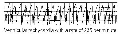
Retrograde ventriculoatrial conduction may occur, which can generate an ECG complex similar to PSVT with aberrant conduction.
Electrolytes, including serum calcium, magnesium, and phosphate levels. Ionised calcium levels are preferred over total serum calcium. Hypokalaemia, hypomagnesaemia, and hypocalcaemia may predispose patients either to conventional VT or torsades de pointes.
Levels of therapeutic drugs - eg, digoxin.
Evaluate for myocardial ischaemia: serum troponin I levels, or other cardiac markers.
CXR: if there is a possibility of congestive heart failure or other cardiopulmonary pathology as contributing factors.
Enfermedades asociadas
Arrhythmia may occur with or without either myocardial ischaemia or infarction.
Accelerated idioventricular rhythm (sometimes termed slow ventricular tachycardia):
Presents with a rate of 60-100 bpm.
Typically occurs with underlying heart disease (coronary or structural).
It is transient, and only rarely is associated with haemodynamic compromise or collapse.
Treatment is usually not required unless there is haemodynamic impairment.
Ventricular tachycardias treatment
Address the ABCs of resuscitation and provide basic life support and advanced life support as necessary, urgent transfer to hospital, venous access, oxygen and ECG rhythm strip monitoring. See advanced life support algorithm.4
Pulseless VT
Is treated as for VF in line with the advanced life support algorithm.4
Unstable VT (reduced cardiac output)
Ver adult tachycardia (with pulse) algorithm.4
VF or pulseless VT is treated by unsynchronised defibrillation; whereas other VTs can be treated with synchronised cardioversion.
Most patients respond to low levels of energy (eg, starting at 50 J biphasic or 100 J monophasic).
Synchronised defibrillation in unstable VT may cause R-on-T deterioration to VF.
Defibrillation is followed by airway management if required, supplemental oxygen, vascular access, and anti-arrhythmic therapy.
Advanced cardiac life support: amiodarone is often used for haemodynamically unstable VT. Replenishment of magnesium and/or other electrolytes may be a valuable adjunct to anti-arrhythmic therapies.
Stable VT
Stable VT patients do not experience symptoms of haemodynamic decompensation.
Unlike other dysrhythmias, VT tends to deteriorate into unstable states and more malignant dysrhythmias.
Therefore, stable VT should be treated with lidocaine or timely cardioversion if lidocaine is ineffective.
The evidence on percutaneous (non-thoracoscopic) epicardial catheter radiofrequency ablation for VT is limited but has shown that the procedure is effective in carefully selected patients and raises no major safety issues.5
Refractory VT
After the initial dose of amiodarone 300 mg IV, it may be followed by an infusion of 900 mg over 24 hours.4
Desfibriladores cardioversores implantables (DCI)
National Institute for Health and Care Excellence (NICE) guidance recommends that ICDs should be considered for patients in the following categories:6
People with previous serious ventricular arrhythmia - that is, people who, without a treatable cause:
Have survived a cardiac arrest caused by either ventricular tachycardia (VT) or ventricular fibrillation; o
Have spontaneous sustained VT causing syncope or significant haemodynamic compromise; o
Have sustained VT without syncope or cardiac arrest, and also have an associated reduction in left ventricular ejection fraction (LVEF) of 35% or less but their symptoms are no worse than class III of the New York Heart Association (NYHA) functional classification of heart failure.
People who:
Have a familial cardiac condition with a high risk of sudden death, such as long QT syndrome, hypertrophic cardiomyopathy, Brugada syndrome or arrhythmogenic right ventricular dysplasia; o
Have undergone surgical repair of congenital heart disease.
ICDs, cardiac resynchronisation therapy (CRT) with defibrillator (CRT‑D) or CRT with pacing (CRT‑P) may be considered as choices for people with heart failure who have left ventricular dysfunction with an LVEF of 35% or less.
Complications of ventricular tachycardias
Congestive cardiac failure and cardiogenic shock.
VT may deteriorate into VF.
Pronóstico
If treated rapidly, VT generally has a favourable short-term outcome.
Long-term prognosis depends upon the underlying cardiac disease.
Actualizaciones exclusivas para profesionales de la salud
Mantente informado con las últimas actualizaciones clínicas, perspectivas profesionales y orientación basada en evidencia. El boletín de Patient Pro selecciona contenido esencial para profesionales de la salud, entregado directamente en tu bandeja de entrada.
Al suscribirte aceptas nuestros Política de Privacidad. Puedes darte de baja en cualquier momento. Nunca vendemos tus datos.
Lecturas adicionales y referencias
- Whitaker J, Wright MJ, Tedrow U; Diagnosis and management of ventricular tachycardia. Clin Med (Lond). 2023 Sep;23(5):442-448. doi: 10.7861/clinmed.2023-23.5.Cardio3. Epub 2023 Sep 29.
- Cronin EM, Bogun FM, Maury P, et al; 2019 HRS/EHRA/APHRS/LAHRS expert consensus statement on catheter ablation of ventricular arrhythmias. J Arrhythm. 2019 May 10;35(3):323-484. doi: 10.1002/joa3.12185. eCollection 2019 Jun.
- Lopez EM, Malhotra R; Ventricular Tachycardia in Structural Heart Disease. J Innov Card Rhythm Manag. 2019 Aug 15;10(8):3762-3773. doi: 10.19102/icrm.2019.100801. eCollection 2019 Aug.
- Edhouse J, Morris F; ABC of clinical electrocardiography: Broad complex tachycardia - Part II. BMJ. 2002 Mar 30;324(7340):776-9.
- Weisfeldt ML, Everson-Stewart S, Sitlani C, et al; Ventricular tachyarrhythmias after cardiac arrest in public versus at home. N Engl J Med. 2011 Jan 27;364(4):313-21.
- Biblioteca de ECG
- Guías de soporte vital avanzado para adultos 2021; Consejo de Reanimación del Reino Unido
- Ablación por radiofrecuencia con catéter epicárdico percutáneo (no toracoscópico) para taquicardia ventricular; Guía de Procedimientos Intervencionistas de NICE, marzo de 2009
- Desfibriladores cardioversores implantables y terapia de resincronización cardíaca para arritmias e insuficiencia cardíaca; Guía de Evaluación de Tecnología del NICE, junio de 2014
Sobre el autorVer biografía completa
Actualizado por

Dr Colin Tidy, MRCGP
Médico General, Autor Médico
MBBS, MRCGP, MRCP (Paediatrics), DCH
El Dr. Colin Tidy es un médico del NHS, con sede en Oxfordshire.
Acerca del revisorVer biografía completa

Dr Hayley Willacy, FRCGP
Médico General, Autor Médico
MBChB (1992), DRCOG, DFFP, MRCOG (Part 1) MRCGP (2007), DFSRH (2013), MSc - medical education (2020)
La Dra. Hayley Willacy fue una médica general del NHS que trabajaba en el noroeste de Inglaterra, quien se retiró de la práctica clínica en 2022 después de 30 años.
Historial del artículo
Artículo también disponible en Inglés, Alemán, Español, Francés, Italiano, Portugués, Hindi, Hebreo, Árabe, y Sueco.
Siguiente revisión prevista: 15 de octubre de 2027
16 Oct 2024 | Última versión
Acceso sencillo a tratamientos y servicios de salud confiables en línea.
Common conditions
Get help with common conditions through the NHS, without a GP appointment.
 Sinusitis aguda
 Amigdalitis bacteriana
 Impetigo
 Picaduras de insectos
 Herpes zósterInfección del tracto urinario (ITU)
Más sobre enfermedades cardiovasculares
- Síndrome coronario agudo
- Edema pulmonar agudo
- Monitoreo ambulatorio de la presión arterial
- Fármacos antifibrinolíticos y hemostáticos
- Trombosis de la vena axilar
- Enzimas cardíacas y marcadores para infarto de miocardio
- Taponamiento cardíaco
- Dolor torácico de tipo cardíaco en atención primaria
- Eventos cerebrovasculares
- Enfermedad cardíaca congénita en adultos
- Vasoespasmo de la arteria coronaria
- Ecocardiografía
- Hiperlipidemia
- Prevención de enfermedades cardiovasculares
- Enfermedad renovascular
- Prevención del accidente cerebrovascular
- Fenómeno de robo subclavio
- Taquicardia supraventricular en adultos
- Síncope
- Aneurismas de la aorta torácica