Nevos intradérmicos y compuestos
Revisado por Dr Rosalyn Adleman, MRCGPÚltima actualización por Dr Doug McKechnie, MRCGPÚltima actualización 25 de marzo de 2022
Cumple con las directrices editoriales
- DescargarDescargar
- Compartir
- Language
- Discusión
- Versión en audio
- Agregar a fuentes preferidas en Google
Profesionales Médicos
Los artículos de Referencia Profesional están diseñados para ser utilizados por profesionales de la salud. Están escritos por médicos del Reino Unido y se basan en evidencia de investigación, así como en guías del Reino Unido y Europa. Puede encontrar uno de nuestros artículos de salud más útil.
Vea también el separado Lesiones cutáneas pigmentadas artículo.
What are intradermal naevi?1 2
Intradermal naevi are a form of melanocytic naevus. A melanocytic naevus (or 'mole') is a common benign skin lesion due to a local proliferation of pigment cells (melanocytes). A brown or black melanocytic naevus contains the pigment melanin, so may also be called a pigmented naevus.
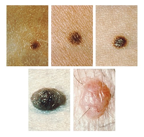
An intradermal naevus has naevus cell nests in the dermis, and presents as a papule, plaque or nodule with a pedunculated, papillomatous or smooth surface.
Some intradermal naevi may have the same degree of pigmentation as the surrounding skin. They are the classical skin or flesh-coloured 'mole', elevated from the skin's surface, that most people would recognise as such. Some are pigmented.3
Intradermal naevi may appear spontaneously or grow from a pre-existing pigmented mole. They usually develop from the end of childhood onwards and may appear at any stage throughout adulthood, although they are probably quite rare as a new phenomenon after the age of 60. Intradermal naevi have the following characteristics:
Small (5 mm-1 cm).
Raised from the surface of the skin (rounded, dome-shaped, pedunculated or warty appearance).
May be associated with hair growth (particularly in older patients).
How common are intradermal naevi? (Epidemiology)4
Intradermal naevi are extremely common, as are all the melanocytic naevi. Indeed they affect so many people that some consider that they cannot be classed as a pathological entity - rather, a normal variant.
Symptoms of intradermal naevi
Intradermal naevi do not present that often, as most people recognise them as a benign dermatological phenomenon.
They may be detected coincidentally during a consultation, or brought up as a 'whilst I'm here' phenomenon.
They are most likely to present if they are newly noticed.
Visual appearance of intradermal naevi
Intradermal naevi
Diagnóstico diferencial5
History and appearance of intradermal naevi are fairly characteristic and so they are not usually confused with other lesions.
They may resemble early carcinoma de células basales o una neurofibroma.
Basal cell carcinoma will usually have a shorter history, be noted to be growing quite quickly and have associated telangiectasia.
Where there is doubt as to the diagnosis then excision biopsy will resolve the question.
Investigaciones
None is usually required.
If there has been significant recent growth of intradermal naevi then consider excision biopsy to exclude basal cell carcinoma.
Where a previously non-pigmented lesion develops pigmentation then excision biopsy should be carried out.
Intradermal naevi treatment
No treatment is required unless the patient is concerned about the lesion's cosmetic appearance or there are suspicions of an alternative diagnosis.
Excision biopsy may be used for diagnostic purposes.
Shave and cautery (dermal electrosurgical shave excision) are a good method for removing them, with apparently better cosmetic results than excision biopsy.6
Injection of local anaesthetic directly into the lesion before shaving may improve the cosmetic result.7
Facial lesions may be best removed for cosmetic purposes by a dermatological surgeon or plastic surgeon, particularly in young patients, due to the high risk of medicolegal claims in this area of practice if there is a poor cosmetic outcome.
Complicaciones y pronóstico
Intradermal naevi have no complications as such and are benign, slow-growing lesions.
If they occur in the external auditory meatus they can obstruct the auditory canal and impair hearing.8
There are potential complications associated with their removal.
Prognosis is excellent, as it is a benign lesion with no risk of transformation to melanoma.
What are compound naevi?2
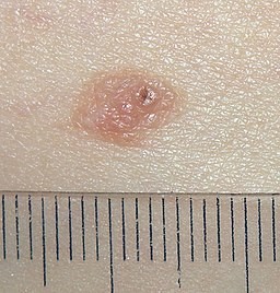
Compound naevi have nests of naevus cells at the epidermal-dermal junction as well as within the dermis, and present as a central raised area surrounded by a flat patch.1
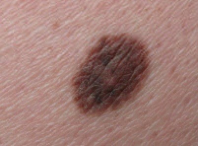
Compound naevi are dark brown in colour. Compound naevi are considered to be benign neoplasms of melanocytes if they arise in later life.9
Compound naevi arise from a flat (junctional) naevus that exists earlier in life and may have a raised central portion of deeper pigmentation with surrounding tan-brown macular pigmentation. Pigmentation may be uneven within the naevus but is usually symmetrically distributed. Compound naevi are usually of a round/oval shape and roughly 2 mm-7 mm in diameter. They may exist with a variable degree of pigmentation and even be the same colour as the surrounding skin. Their name is derived from the fact that they contain junctional melanocytes (responsible for their pigmentation) and intradermal melanocytes (responsible for the elevation of the lesion).
How common are compound naevi? (Epidemiology)
Melanocytic naevi in the general population are exceedingly common in congenital and acquired form. Their prevalence is so high that some believe they cannot even be considered an abnormality or pathological entity, as most people with light-coloured skin will have at least a few.10
Compound naevi are much more common in ethnic groups with light skin but they still have an appreciable prevalence in those with more pigmented skin. Congenital melanocytic naevi occur in approximately 1% of newborns at birth. Acquired melanocytic naevi are usually seen from the age of one year, peak in number during the second and third decades of life and disappear between the seventh to ninth decades.11
Signs and symptoms of compound naevi (presentation)
Compound naevi symptoms
Establish if the lesion is congenital or acquired (compound naevi are acquired).
Cuando una lesión se presenta médicamente, es importante determinar si ha habido algún síntoma asociado, como:
Ampliación.
Cambio en forma o tamaño.
Cambio en la pigmentación.
Picazón/dolor/irritación.
Sangrado.
Compound naevi signs
Examine la lesión con luz brillante, preferiblemente luz del día si está disponible.
Use drawings or photography to note the site(s), size or shape and pigmentation of the lesion.
Establish that the lesion has the typical pattern of pigmentation and is raised from the level of the skin.
Distinguish from other similar raised benign tumours of the skin:
Dermatofibromas tend to feel firm or hard on palpation, whereas compound naevi are softer.
Seborrhoeic warts tend to have a 'stuck-on' appearance, rather than blending into the surrounding skin.
El melanoma tiende a ser más oscuro, tener un borde irregular, ser asimétrico y haber crecido recientemente.
Visual appearance of compound naevi
Compound naevus with size gauge
Compound naevus on left buttock
Diagnóstico diferencial5
Léntigos.
Lunar atípico (nevus displásico).
Otros nevos melanocíticos.
Granuloma piógeno (usually reddish but may be brown).
Seborrhoeic keratosis.
Acanthoma.
Histiocytoma.
Skin tag (acrochordon).
Neuroma.
Nevus de Ota e Ito.
Diagnóstico compound naevi (investigations)
No investigations are indicated in the case of a common, acquired compound naevus that has not undergone any recent change.
Algunos dermatólogos pueden usar la dermatoscopia para intentar distinguir la naturaleza de las lesiones pigmentadas.12
Si hay alguna sospecha de melanoma maligno, la investigación de elección es la biopsia por escisión.
Compound naevi treatment
If the diagnosis of compound naevus is clear and there has been no change in a long-standing lesion then reassurance and monitoring of the lesion are all that is usually required.
Where there is any doubt as to the diagnosis of compound naevi, perform excision biopsy or refer for dermatological advice.
Realizar una biopsia por escisión siempre que la lesión presente:
Crecido.
Presentar síntomas.
Asimetría desarrollada.
Desarrolló un borde irregular.
Alteró su grado o patrón de pigmentación.
Lesiones satélite desarrolladas.
Complicaciones y pronóstico
Compound naevi are benign lesions. They do not cause complications and they have an excellent prognosis.
Actualizaciones exclusivas para profesionales de la salud
Mantente informado con las últimas actualizaciones clínicas, perspectivas profesionales y orientación basada en evidencia. El boletín de Patient Pro selecciona contenido esencial para profesionales de la salud, entregado directamente en tu bandeja de entrada.
Al suscribirte aceptas nuestros Política de Privacidad. Puedes darte de baja en cualquier momento. Nunca vendemos tus datos.
Lecturas adicionales y referencias
- Mejorando los resultados para personas con tumores de piel, incluyendo melanoma; Guía NICE (actualización de mayo de 2010)
- Almazan-Fernandez FM, Fernandez-Pugnaire MA, Hernandez-Gil J, et al; Homogeneous blue pattern: A rare presentation in an acral congenital melanocytic nevus. Dermatol Online J. 2010 Aug 15;16(8):10.
- Ferreira L, Jham B, Assi R, et al; Oral melanocytic nevi: a clinicopathologic study of 100 cases. Oral Surg Oral Med Oral Pathol Oral Radiol. 2015 Sep;120(3):358-67. doi: 10.1016/j.oooo.2015.05.008. Epub 2015 May 28.
- Nevus melanocítico; DermNet NZ
- Damsky WE, Bosenberg M; Nevos melanocíticos y melanoma: desentrañando una relación compleja. Oncogene. 19 de octubre de 2017;36(42):5771-5792. doi: 10.1038/onc.2017.189. Publicado en línea el 12 de junio de 2017.
- Suwattee P, Schram SE, Warshaw EM; Digital polarized light dermoscopy of clinically nonpigmented dermal nevi. Dermatol Surg. 2007 Sep;33(9):1120-5. doi: 10.1111/j.1524-4725.2007.33229.x.
- Intradermal nevus; Libre Pathology, 2014
- Plensdorf S, Livieratos M, Dada N; Trastornos de Pigmentación: Diagnóstico y Manejo. Am Fam Physician. 15 de diciembre de 2017;96(12):797-804.
- Sardana K, Chakravarty P, Goel K; Manejo óptimo de los nevos melanocíticos adquiridos comunes (lunares): perspectivas actuales. Clin Cosmet Investig Dermatol. 19 de marzo de 2014;7:89-103. doi: 10.2147/CCID.S57782. eCollection 2014.
- Zaiac M, Shah VV, Mlacker S, et al; Local anesthesia injection technique for aesthetic removal of dermal nevi. J Cosmet Dermatol. 2016 Jun 22. doi: 10.1111/jocd.12240.
- Kazikdas KC, Onal K, Kuehnel TS, et al; An intradermal nevus of the external auditory meatus. Eur Arch Otorhinolaryngol. 2006 Mar;263(3):253-5. Epub 2005 Jul 13.
- Trozak D et al; Dermatology Skills for Primary Care: An Illustrated Guide, 2007.
- Compound Nevus; Libre Pathology, 2014
- Russak J et al; Pigmented Lesions in Children, Semin Plast Surg 20(3): 169–179, 2006.
- Kim JK, Nelson KC; Dermoscopic features of common nevi: a review. G Ital Dermatol Venereol. 2012 Apr;147(2):141-8.
Sobre el autorVer biografía completa
Actualizado por

Dr Doug McKechnie, MRCGP
Redactor Médico
MA, MBBS, MSc, DRCOG, MRCP(UK), MRCGP(2021), FHEA
El Dr. Doug McKechnie es un médico de cabecera del NHS que trabaja en Londres. Trabaja a tiempo completo en la práctica clínica y también es el Subdirector del módulo de Práctica Clínica y Profesional en la Escuela de Medicina del University College London.
Acerca del revisorVer biografía completa
Dr Rosalyn Adleman, MRCGP
MRCGP
La Dra. Rosalyn Adleman es una médica de cabecera del NHS que trabaja en el norte de Londres.

Pregunta, comparte, conecta.
Navega por discusiones, haz preguntas y comparte experiencias en cientos de temas de salud.

¿Te sientes mal?
Evalúa tus síntomas en línea de forma gratuita
Más en dermatología
- verrugas anogenitales
- Tratamiento a ciegas de infección bacteriana
- Dermatitis herpetiforme
- Dermatofibroma
- Herpes genital durante el embarazo
- Enfermedad del gusano de Guinea
- Hidradenitis supurativa
- Impetigo
- Nevus de unión
- Síndrome de lipodistrofia
- Esclerodermia localizada
- Melanoma maligno de la piel
- Miositis - polimiositis y dermatomiositis
- Porfirias
- Úlceras por presión
- Piojos púbicos y corporales
- PUVA
- Enfermedad de Schamberg
- Toxocariasis
- Xantelasma