Escaneo DEXA
Revisado por pares por Dr Hayley Willacy, FRCGP Última actualización por Dr Colin Tidy, MRCGPÚltima actualización 20 Mar 2023
Cumple con las directrices editoriales
- DescargarDescargar
- Compartir
- Language
- Discusión
- Versión en audio
- Agregar a fuentes preferidas en Google
En esta serie:OsteoporosisBisfosfonatosAlimentos ricos en calcioDeficiencia de vitamina DPrevención de la osteoporosis inducida por esteroides
DEXA scans (also called DXA scans or bone density scans) are used to check the density of bones. This test uses X-rays to show how strong bones are. A DEXA scan is different from a escaneo óseo, which used radioactive chemicals to create a picture of the bones.
De un vistazo
Un escaneo DEXA mide la densidad ósea utilizando rayos X de baja energía.
Los huesos densos son más fuertes y menos propensos a romperse.
La exploración generalmente toma de 5 a 20 minutos mientras estás acostado en un sofá.
Las exploraciones DEXA utilizan un nivel muy bajo de radiación de rayos X.
No necesitas preparación especial; evita la ropa con metal.
Puede ser aconsejable si ha tenido una fractura o está en riesgo de tener huesos 'frágiles'.
Las exploraciones DEXA no se recomiendan para mujeres que están embarazadas.
Nota: la información a continuación es solo una guía general. Los arreglos y la forma en que se realizan las pruebas pueden variar entre diferentes hospitales. Siempre siga las instrucciones dadas por su médico o hospital local.
What is a DEXA scan?
DEXA stands for 'dual-energy X-ray absorptiometry'. DEXA (also sometimes known as DXA) is a test that measures the density of bones. Density means how much of something there is in a certain amount of space. The denser the tissue, the less X-rays pass through.
Air and water are less dense than solid things such as bone. This is because the particles which make air and water are not held closely together. In general, the more dense the bone, the stronger it is, and the less likely it is to break (fracture).
There are two different types of DEXA scanning devices:
Central DEXA devices are large machines that can measure bone density in the centre of your skeleton, such as your hip and spine.
Peripheral DEXA devices are smaller, portable machines that are used to measure bone density on the periphery of your skeleton, such as your wrist, heel or finger. These are mainly to get an idea about whether further tests are needed, as they are not as accurate as the larger DEXA machines.
How does a DEXA scan work?
A DEXA scan uses low-energy X-rays. A machine sends X-rays from two different sources through the bone being tested. Bone blocks a certain amount of the X-rays. The more dense the bone is, the less X-rays get through to the detector. By using two different X-ray sources rather than one it greatly improves the accuracy in measuring the bone density.
The amount of X-rays that comes through the bone from each of the two X-ray sources is measured by a detector. This information is sent to a computer which calculates a score of the average density of the bone. A low score indicates that the bone is less dense than it should be, some material of the bone has been lost and it is more prone to fracture.
How is a DEXA scan done?
You lie on your back on a couch and are asked to keep still while an X-ray detector (the 'scanner') comes over the area to be tested. An X-ray machine fires X-rays towards the detector. The bones commonly scanned are the bones of the back (the vertebrae), hip and wrist.
Smaller peripheral scanners are available in some places and can be used to check the bone mass density of the heel, wrist or finger.
How long does a DEXA scan take?
The scan usually takes between 5 and 20 minutes, depending on which part of your body is being examined and whether a central or peripheral scanner is being used. There is no 'tunnel' to pass through as there is in other types of scans such as an MRI or CT scan, so it should not affect people who do not like being in enclosed spaces.
DXA scanner
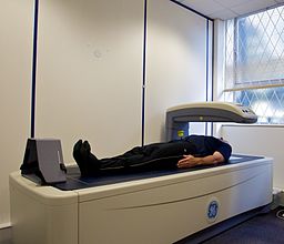
© Nick Smith photography (ALSPAC website), via Wikimedia Commons
Are DEXA scans safe?
DEXA scans use a very low level of X-ray radiation. This means it is safe for the technician doing the scan to stay in the room with you. (In standard X-ray tests, the technician has to stay behind a protective screen.)
Preparing for a DEXA scan
You do not need to do any special preparation prior to a DEXA scan. You can normally remain fully clothed, although you will need to avoid or remove clothes with metal in them (for example, zips, belts, buttons). You may also be asked to remove jewellery for the scan. In some places, you may be given a gown to wear.
Who should have a DEXA scan?
A DEXA scan may be advised if you have had a fracture of a bone after a minor injury. It may also be advised if you are considered at increased risk of 'thinning' of the bones (osteoporosis) and therefore at increased risk of having a fracture in future.
If your doctor thinks you have risk factors for osteoporosis, they may use a risk calculator such as one called FRAX® or QFracture®. This gives an idea of how likely you are to fracture your bones after a minor knock. If your risk is at a medium level, your doctor would then arrange a DEXA scan. This enables them to gain a clearer picture of your risk and then to decide whether you need any treatment.
DEXA scans are also used to monitor whether treatment for osteoporosis is working.
DEXA scans are not advised for women who are pregnant. You should also not have a DEXA scan within two weeks of certain other types of scans - for example, those using contrast dye.
For further information, see the separate leaflet called Osteoporosis.
Selecciones del paciente para Imágenes

Pruebas e investigaciones
Pruebas de bario
Las pruebas de bario se utilizan para ayudar a ver el contorno de varias partes del intestino (tracto gastrointestinal). Estas incluyen el esófago, el estómago, el intestino delgado y el colon (intestino grueso). Las pruebas de rayos X con bario se realizan con menos frecuencia en la actualidad. Hoy en día, generalmente examinamos el intestino con un telescopio flexible (endoscopia o colonoscopia). Sin embargo, todavía hay un lugar para las pruebas de bario para ayudar a evaluar varios problemas del intestino. Nota: la información a continuación es solo una guía general. Los arreglos y la forma en que se realizan las pruebas pueden variar entre diferentes hospitales. Siempre siga las instrucciones dadas por su médico o hospital local.
por la Dra. Rosalyn Adleman, MRCGP

Pruebas e investigaciones
prueba de rayos X
Las pruebas de rayos X muestran huesos y ciertos otros tejidos.
por la Dra. Hayley Willacy, FRCGP
Preguntas frecuentes
¿Cuál es el propósito de una exploración DEXA?
Una exploración DEXA se utiliza para medir la densidad de los huesos. Esta medición ayuda a determinar cuán fuertes son tus huesos y cuán probable es que se rompan (fracturen). Puede recomendarse si ya has tenido una fractura por una lesión menor, o si se te considera en alto riesgo de osteoporosis (adelgazamiento de los huesos).
¿Una prueba de densidad ósea muestra cáncer?
El artículo afirma que una exploración DEXA mide la densidad ósea para evaluar el riesgo de fracturas o monitorear el tratamiento de la osteoporosis. No menciona que una exploración DEXA se utilice para detectar cáncer.
¿Necesito hacer algo especial para prepararme para una exploración DEXA?
Normalmente no necesitas ninguna preparación especial. Generalmente puedes permanecer completamente vestido, pero deberás evitar o quitarte cualquier prenda con metal, como cremalleras, cinturones o botones. También se te podría pedir que te quites las joyas, y en algunos casos, se te podría dar una bata para usar.
¿Quién no debería hacerse una exploración DEXA?
Las exploraciones DEXA no se recomiendan para mujeres embarazadas. Además, no deberías someterte a una exploración DEXA dentro de las dos semanas posteriores a haber tenido otros tipos de exploraciones que impliquen el uso de tinte de contraste.
¿Cuál es la diferencia entre los dispositivos DEXA centrales y periféricos?
Los dispositivos DEXA centrales son máquinas grandes que miden la densidad ósea en las partes centrales de tu esqueleto, como la cadera y la columna vertebral. Los dispositivos DEXA periféricos son máquinas más pequeñas y portátiles que se utilizan para áreas como la muñeca, el talón o el dedo. Los dispositivos periféricos principalmente indican si se necesita realizar más pruebas, ya que no son tan precisos como las máquinas centrales más grandes.
Lecturas adicionales y referencias
- Osteoporosis: evaluando el riesgo de fractura por fragilidad; Guía Clínica NICE (agosto 2012, actualizada en febrero 2017)
- Management of osteoporosis and the prevention of fragility fractures - A national clinical guideline; Scottish Intercollegiate Guidelines Network (SIGN - January 2021)
- Osteoporosis - prevention of fragility fractures; NICE CKS, julio 2021 (acceso solo en el Reino Unido)
- Clinical guideline for the prevention and treatment of osteoporosis; National Osteoporosis Guideline Group (updated September 2021)
Sobre el autorVer biografía completa

Dr Colin Tidy, MRCGP
Médico General, Autor Médico
MBBS, MRCGP, MRCP (Paediatrics), DCH
El Dr. Colin Tidy es un médico del NHS, con sede en Oxfordshire.
Acerca del revisorVer biografía completa

Dr Hayley Willacy, FRCGP
Médico General, Autor Médico
MBChB (1992), DRCOG, DFFP, MRCOG (Part 1) MRCGP (2007), DFSRH (2013), MSc - medical education (2020)
La Dra. Hayley Willacy fue una médica general del NHS que trabajaba en el noroeste de Inglaterra, quien se retiró de la práctica clínica en 2022 después de 30 años.
Historial del artículo
La información en esta página está escrita y revisada por pares por clínicos calificados.
Artículo también disponible en Inglés, Alemán, Español, Francés, Italiano, Portugués, Hindi, Hebreo, Árabe, y Sueco.
Próxima revisión: 18 Mar 2028
20 Mar 2023 | Última versión

Pregunta, comparte, conecta.
Navega por discusiones, haz preguntas y comparte experiencias en cientos de temas de salud.

¿Te sientes mal?
Evalúa tus síntomas en línea de forma gratuita
Suscríbete al boletín de Patient
Tu dosis semanal de consejos de salud claros y confiables, escritos para ayudarte a sentirte informado, seguro y en control.
Al suscribirte aceptas nuestros Política de Privacidad. Puedes darte de baja en cualquier momento. Nunca vendemos tus datos.
Más en pruebas e investigaciones
- Enema de bario
- Grupos y tipos sanguíneos
- Análisis de sangre para detectar inflamación
- Detección cervical
- Cistoscopia
- Ecocardiograma
- Hemograma completo y frotis de sangre
- Escaneo de galio
- Pruebas de audición
- Histerosalpingografía
- Exámenes físicos de recién nacidos
- prueba de detección en recién nacidos
- Osmolalidad, osmolaridad y homeostasis de líquidos
- Espirometría
- Pruebas de ITS
- Prueba de sudor
- Talasemia
- Escaneos de tiroides y pruebas de captación
- Ecografía
- Cistouretrograma de vaciamiento