Verruga seborreica
Revisado por pares por Dra. Toni Hazell, MRCGPÚltima actualización por Dr Doug McKechnie, MRCGPÚltima actualización 17 de febrero de 2025
Cumple con las directrices editoriales
- DescargarDescargar
- Compartir
- Language
- Discusión
- Versión en audio
- Agregar a fuentes preferidas en Google
Profesionales Médicos
Los artículos de Referencia Profesional están diseñados para ser utilizados por profesionales de la salud. Están escritos por médicos del Reino Unido y se basan en evidencia de investigación, así como en guías del Reino Unido y Europa. Puede encontrar el Verrugas seborréicas artículo más útil, o uno de nuestros otros artículos de salud.
Synonyms: seborrhoeic keratosis, basal cell papilloma
What are seborrhoeic warts?
Seborrhoeic warts (also known as seborrhoeic keratoses) are common benign, hyperkeratotic skin lesions associated with ageing.
Causes of seborrhoeic warts (aetiology)1
The cause is not fully understood. Ageing is closely linked to the development of seborrhoeic keratoses. Sunlight and UV light exposure appears to play a role in causation, given the typical distribution of seborrhoeic warts. They have been known to follow sunburn.
An infectious aetiology has been proposed, with human papillomavirus and Merkel cell polyomavirus detected in seborrhoeic warts in some studies. However, these were felt to either be surface contamination or non-causal co-infection.1
Some cases of multiple seborrhoeic warts are inherited in an autosomal dominant pattern.2 Oncogenic mutations are involved in pathogenesis and a broad spectrum of somatic mutations in the FGFR3, PIK3CA, RAS, AKT1 and EGFR genes has been noted.3
How common are seborrhoeic warts? (Epidemiology)45
Presence and frequency increases with age: almost all elderly patients have some. It has been estimated that over 90% of adults aged 60 or more have at least one seborrhoeic wart.
They usually present from the fourth decade onwards.
No sex difference exists.
Seborrhoeic warts are less common in people with darker skin tones, but can occur in anyone.
The trunk and face are the sites most commonly affected.
It is common to have more than one lesion and there may be many lesions.
Symptoms of seborrhoeic warts (presentation)
Typical clinical appearance
Flat-topped or warty-looking lesions that appear to be 'stuck on' to the skin.
Usually pigmented, sometimes deeply and may even be black. Others can be paler in colour.
There is usually a well-circumscribed border.
Size ranges from 1 mm to several cm.
The surface is usually pitted and irregular with visible keratin dots giving a granular and rough appearance.
Initially, lesions are velvety and soft in texture, before developing a warty surface and becoming uneven, with multiple plugged follicles.
The surface may become covered by adherent greasy scale.
Multiple lesions may align along skin folds.
They are usually asymptomatic but may become irritated, itchy or inflamed spontaneously or after minor trauma.
Black pigmented verrucous lesion
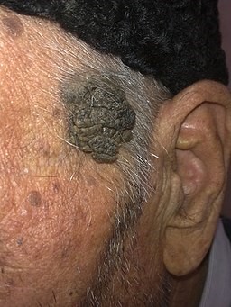
© Alborz Fallah, CC BY-SA 3.0, vía Wikimedia Commons
Queratosis seborreica
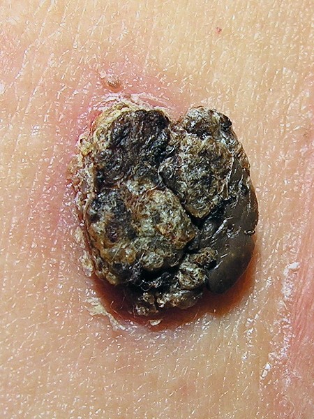
© Lmbuga, Public domain, via Wikimedia Commons
Características dermoscópicas56
Thickened epidermis with multiple milia-like cysts and comedo-like openings.
Gyri and sulci (looking like the surface of the brain).
Fingerprint-like structures.
Bloods vessels are fine, regular and hairpin in shape and surrounded by a milky halo.
'Moth-eaten' borders in the thinner lesions.
Variants
Less common variants of seborrhoeic warts include:
Stucco keratoses - multiple skin-coloured or white, dry, scaly lesions often seen on the extremities (dorsa of hands, forearms, ankles and feet).7
Dermatosis papulosa nigra - multiple small, brown or black pedunculated lesions seen on the face of dark-skinned individuals. Often have an earlier onset than typical seborrhoeic warts.8
Solar lentigo - well-circumscribed, flat pigmented patches which occur in sun-exposed areas.9
Melanoacanthoma - very deeply pigmented seborrhoeic warts.10
Diagnóstico diferencial11
Verruca vulgaris.
Condyloma acuminatum.
Fibroepithelial polyp.
Melanocytic naevus.
Pigmented carcinoma de células basales.
Enfermedades asociadas
Although skin malignancy is thought to occur by chance in patients with seborrhoeic warts, studies report the occasional association with Bowen's disease and squamous epithelial dysplasia.12 This occurs more frequently in regions with high solar ultraviolet levels.13
Rarely, a sudden onset or increase in the number of seborrhoeic warts can herald an underlying malignancy (usually adenocarcinoma of the stomach but also colon, breast and lung).14 It can be associated with acanthosis nigricans. This sudden increase is known as the Leser-Trélat sign.5 The same phenomenon without internal malignancy is known as a pseudo-Leser-Trélat sign.15
Management of seborrhoeic warts5
Reassurance: most often, no treatment is required.
Remove where there is cosmetic dislike, repeated irritation or chafing from clothes, or diagnostic uncertainty.
Removal by cryotherapy (this may require repeat treatments), curettage and cautery or shave excision are effective and produce a better result than excision and suture, although a pale white scar can be left.
Dermoscopy may be used by appropriately trained GPs to assist in diagnosis.16
Cuándo derivar
Usually, they can be managed in primary care. However, patients with lesions requiring removal from difficult areas should be referred. Lesions that are suspicious of melanoma (either with the naked eye or by dermoscopy) should be sent as a cancer network two-week referral to a dermatologist.16
Complications of seborrhoeic warts
Repeated irritation and inflammation where lesions catch on clothing.
Aesthetic dislike.
Concerns regarding malignancy:
It is harder to notice a malignant melanoma arise amongst multiple seborrhoeic warts.
Rarely, melanoma in situ can arise within a seborrhoeic wart, although this is rare.17
Pronóstico
Although seborrhoeic warts are benign, they do not spontaneously resolve and they become larger and thicker with time.
Actualizaciones exclusivas para profesionales de la salud
Mantente informado con las últimas actualizaciones clínicas, perspectivas profesionales y orientación basada en evidencia. El boletín de Patient Pro selecciona contenido esencial para profesionales de la salud, entregado directamente en tu bandeja de entrada.
Al suscribirte aceptas nuestros Política de Privacidad. Puedes darte de baja en cualquier momento. Nunca vendemos tus datos.
Lecturas adicionales y referencias
- Noiles K, Vender R; Are all seborrheic keratoses benign? Review of the typical lesion and its variants. J Cutan Med Surg. 2008 Sep-Oct;12(5):203-10.
- Gorai S, Ahmad S, Raza SSM, et al; Update of pathophysiology and treatment options of seborrheic keratosis. Dermatol Ther. 2022 Dec;35(12):e15934. doi: 10.1111/dth.15934. Epub 2022 Nov 1.
- Seborrheic Keratosis; Herencia Mendeliana en Línea en el Hombre (OMIM)
- Hafner C, Hafner H, Groesser L; Genetic basis of seborrheic keratosis and epidermal nevi. Pathologe. 2014 Sep;35(5):413-23. doi: 10.1007/s00292-014-1928-9.
- Seborrhoeic Keratoses; DermNet NZ
- Queratosis seborreica; Sociedad de Dermatología de Atención Primaria
- Marghoob AA, Usatine RP, Jaimes N; Dermoscopy for the family physician. Am Fam Physician. 2013 Oct 1;88(7):441-50.
- Stucco keratosis. Primary Care Dermatology Society. Accessed 4th Jan 2025.
- Dermatosis Papulosa Nigra. StatPearls.
- Solar lentigo; DermNet NZ
- Vasani RJ, Khatu SS; Melanoacanthoma: Uncommon presentation of an uncommon condition. Indian Dermatol Online J. 2013 Apr;4(2):119-21. doi: 10.4103/2229-5178.110638.
- Seborrheic Keratosis; DermIS (Sistema de Información de Dermatología)
- Rajabi P, Adibi N, Nematollahi P, et al; Bowenoid transformation in seborrheic keratosis: A retrospective analysis of 429 patients. J Res Med Sci. 2012 Mar;17(3):217-21.
- Gaffney DC, Muir JB, De'ambrosis B; Malignant change in seborrhoeic keratoses in a region with high solar ultraviolet levels. Australas J Dermatol. 2013 Apr 10. doi: 10.1111/ajd.12035.
- Ponti G, Luppi G, Losi L, et al; Leser-Trelat syndrome in patients affected by six multiple metachronous primitive cancers. J Hematol Oncol. 2010 Jan 11;3:2.
- Husain Z, Ho JK, Hantash BM; Sign and pseudo-sign of Leser-Trelat: case reports and a review of the literature. J Drugs Dermatol. 2013 May;12(5):e79-87.
- Dermoscopy - an overview; Primary Care Dermatology Society, 2013
- Repertinger S, Wang J, Adickes E, et al; Melanoma in-situ arising in seborrheic keratosis: a case report. Cases J. 2008 Oct 23;1(1):263.
Sobre el autorVer biografía completa

Dr Doug McKechnie, MRCGP
Redactor Médico
MA, MBBS, MSc, DRCOG, MRCP(UK), MRCGP(2021), FHEA
El Dr. Doug McKechnie es un médico de cabecera del NHS que trabaja en Londres. Trabaja a tiempo completo en la práctica clínica y también es el Subdirector del módulo de Práctica Clínica y Profesional en la Escuela de Medicina del University College London.
Acerca del revisorVer biografía completa

Dra. Toni Hazell, MRCGP
MBBS, BSc, MRCGP, DFSRH, Dip GU med, DRCOG, DCH (London, UK, 2000)
La Dra. Toni Hazell se graduó de la Escuela de Medicina del Hospital St. Mary y realizó su VTS en el Hospital Northwick Park.
Historial del artículo
La información en esta página está escrita y revisada por pares por clínicos calificados.
Artículo también disponible en Inglés, Alemán, Español, Francés, Italiano, Portugués, Hindi, Hebreo, Árabe, y Sueco.
Siguiente revisión prevista: 16 de febrero de 2028
17 de febrero de 2025 | Última versión

Pregunta, comparte, conecta.
Navega por discusiones, haz preguntas y comparte experiencias en cientos de temas de salud.

¿Te sientes mal?
Evalúa tus síntomas en línea de forma gratuita
Más en dermatología
- Carcinoma de células basales
- Tratamiento a ciegas de infección bacteriana
- Pie diabético
- Erythema chronicum migrans
- Gota
- Queratoacantoma
- Liquen plano
- Sarampión
- Enfermedad mixta del tejido conectivo
- Infecciones cutáneas por micobacterias
- Necrobiosis lipoidica
- Ameba de vida libre patógena
- Pelagra
- Artritis psoriásica
- Enfermedad ungueal psoriásica
- Vena en araña
- Esclerosis sistémica
- Dedos de los pies y de las manos en palillo
- Venas varicosas