Léntigo
Revisado por pares por Dr Jo SealeÚltima actualización por Dr Toni Hazell, FRCGPÚltima actualización 22 Nov 2022
Cumple con las directrices editoriales
- DescargarDescargar
- Compartir
- Language
- Discusión
- Versión en audio
- Agregar a fuentes preferidas en Google
Profesionales Médicos
Los artículos de Referencia Profesional están diseñados para ser utilizados por profesionales de la salud. Están escritos por médicos del Reino Unido y se basan en evidencia de investigación, así como en guías del Reino Unido y Europa. Puede encontrar uno de nuestros artículos de salud más útil.
What is a lentigo?
Lentigines (plural of lentigo) are flat brown lesions which do not darken following sun exposure (thus differentiating them from ephelides, or true freckles).
Apariencia1
One study of white women found that lentigines were signs of photo-damage whereas there was a genetic component in true freckles.2 They may be any size from 5-20 mm and may be irregular in shape. They occur over the shoulders in young people, especially those who have had a lot of sun exposure and in the elderly on the sun-exposed sites such as the dorsum of the hands and forearms, the face and the neck.
The histopathology may include hyperplasia of the epidermis and pigmentation of the basal layer. A number of different types are found:
Lentigo simplex - the most common type, mainly seen in children and not associated with sun exposure. The spots are 5-15 mm in diameter.
Léntigo solar - related to sun exposure. Seen on skin normally exposed to sunlight (eg, face, hands), they are benign lesions, <5 mm in diameter but may merge to form larger spots. Former peak age of 30-50 is becoming younger with increasing sun exposure. The colour may vary from yellow-brown to black.
Léntigo solar
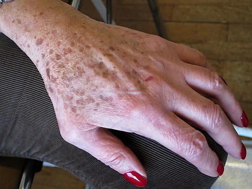
© Alain Gérard, CC BY-SA 4.0, via Wikimedia Commons
Ink spot lentigo - this lesion, so called because it resembles a spot of ink, is a benign lesion limited to sun-exposed areas. They are usually solitary lesions and may be mistaken for melanomas.
PUVA lentigo - these brown macules can appear six months or more after the start of psoralen combined with ultraviolet A (PUVA) treatment for psoriasis in areas exposed to treatment. The macules are usually 3-8 mm in diameter and persist for 3-6 months after treatment has finished. There is also a larger stellate form (which can be up to 3 cm in diameter) which may persist for two years or more.
Radiation lentigo - this appears after a single large exposure to radiation (of Chernobyl proportions rather than post-radiotherapy). There may be other histological signs of radiation damage, such as epidermal atrophy and subcutaneous fibrosis. The malignant potential for such lesions is not known.
Sunbed lentigo - lentigines can appear rapidly after intense exposure, or they may appear after long-term use. They are 2-5 mm in diameter and are most common on the anterior aspects of the arms and legs.
Labial and oral melanotic macules - labial macules usually appear as single lesions on the vermillion of the lower lip. Oral lesions occur on the gingival and buccal mucosa, palate and tongue.
Genital lentigo - in men, this can present as a tan to dark-brown macule anywhere on the glans, shaft or corona. They can achieve a size of 15 mm. In women, lentigines can occur anywhere on the genital mucosa and are usually between 5-15 mm in size.
Lentigines profusa - in this condition, there are widespread lentigines over the arms, legs and genitalia. There are no trigger factors or associated abnormalities (thus differentiating them from a number of lentigo-associated syndromes - see below). The lesions can be 5 mm-2 cm in diameter. The appearance is of dark brown or black freckles but with a more generalised distribution.
Agminated lentiginosis - in this condition, which can be associated with a number of childhood diseases, lentigines appear in a sharply demarcated distribution which often follows the outline of dermatomes. They are tan or dark brown in colour and may be present at birth or develop in early childhood.
A number of lentigo-associated conditions can occur:
LEOPARD - lentigines, electrocardiographic conduction defects, ocular hypertelorism, pulmonary stenosis, abnormal genitalia, restriction of growth, deafness.
Peutz-Jeghers syndrome - gastrointestinal polyps and pigmented macules.
Laugier-Hunziker syndrome - a variable number of pigmented macules appearing commonly around the oral mucosa or lower lip and in other areas.
Myxoma syndromes - a group of disorders including:
LAMB - lentigines, atrial myxomas, mucocutaneous myxomas and blue naevi.
NAME - naevi, atrial myxoma, myxoid neurofibroma and ephelides.
Carney's syndrome - cardiac, cutaneous and mammary myxomatous masses; lentigines; blue naevi; endocrine disorders; testicular tumours.
Diagnóstico diferencial3
Ephelides (freckles) - become darker after sun exposure.
Lentigo maligna - seen mainly on the sun-exposed areas of the face and neck in the elderly; it is slow-growing and sometimes grows to a size of several centimetres. Their size and site differentiates them from lentigines. Lentigo maligna is a pre-cancerous condition. Conversion to a lentigo maligna melanoma can take from a few months to up to 15 years. Transformation to malignancy is lower than in other forms of melanoma, occurring in approximately 5% of patients. However, the risk is higher in larger lesions with a risk of 50% in lesions larger than 4 cm. Identifying lesions that require referral is not easy but worrying signs include changes in size or colour, itching, burning, bleeding, or pain.4 The ABCDE rule of melanoma may be helpful:
Asymmetry.
Sorder irregularity.
Colour variegation.
Diameter greater than 6 mm (the end of a pencil head), although melanoma can occur in lesions less than 6 mm.
Enlargement.
Lentigo maligna
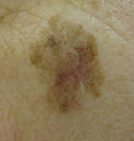
© kilbad, CC BY 3.0, via Wikimedia Commons
Ver el separado Malignant Melanoma of Skin artículo.
Investigaciones5
Biopsy and histology may be used to differentiate the various types of lentigines.
A dermatoscope is occasionally used in the diagnosis of solar lentigo.
The diagnosis of lentigo maligna has been improved by the use of reflectance confocal microscopy (a non-invasive imaging technique that enables in vivo visualisation of the epidermis down to the papillary dermis) and immunohistochemical stains.
Primary care management of lentigo
Unsightly lesions of the face can be lightly frozen, which often improves the cosmetic result.
Tretinoin is occasionally employed to lighten lesions (unlicensed use).6
Any treatment which has a purely cosmetic aim may not be available on the NHS.
Secondary care management of lentigo
Cryotherapy can be used for isolated lentigines. One study found it was more effective than 40% trichloracetic acid for the treatment of solar lentigines although the difference was not significant.
Lasers are useful for a variety of lentigines. Aggressive therapy for using quality-switched lasers is effective in the treatment of solar lentigines but carries the risk of post-inflammatory hyperpigmentation (PIH). For darker skin types, less intensive irradiation reduces this risk, with no reduction in efficacy.7
Intense pulsed light (IPL) is another option.
Cuándo derivar
For doubt over diagnosis and for diagnostic biopsy.
When treatment is required but cannot be provided within primary care - eg, treatment with lasers (Q-switched Nd:YAG or ruby) are effective when available.8
Pronóstico
Lentigines tend to worsen over time but do not become malignant.
Lentigo prevention1
New lesions can be prevented to some extent by sun avoidance. Clothing is more effective than sunscreens.
Avoiding the excessive use of sunbeds helps to prevent tanning-bed lentigines.
Avoidance of a large single dose of ionising radiation helps to prevent radiation lentigines.
Actualizaciones exclusivas para profesionales de la salud
Mantente informado con las últimas actualizaciones clínicas, perspectivas profesionales y orientación basada en evidencia. El boletín de Patient Pro selecciona contenido esencial para profesionales de la salud, entregado directamente en tu bandeja de entrada.
Al suscribirte aceptas nuestros Política de Privacidad. Puedes darte de baja en cualquier momento. Nunca vendemos tus datos.
Lecturas adicionales y referencias
- Goncharova Y, Attia EA, Souid K, et al; Dermoscopic features of facial pigmented skin lesions. ISRN Dermatol. 2013;2013:546813. doi: 10.1155/2013/546813. Epub 2013 Feb 3.
- Léntigo; DermNet NZ
- Ezzedine K, Mauger E, Latreille J, et al; Freckles and solar lentigines have different risk factors in Caucasian women. J Eur Acad Dermatol Venereol. 2012 Aug 28. doi: 10.1111/j.1468-3083.2012.04685.x.
- Barkal C et al; Clinical Atlas of Skin Tumors, 2014
- Lentigo maligna and lentigo maligna melanoma; DermNet NZ
- Kasprzak JM, Xu YG; Diagnosis and management of lentigo maligna: a review. Drugs Context. 2015 May 29;4:212281. doi: 10.7573/dic.212281. eCollection 2015.
- Kircik LH; Safety and efficacy evaluation of tretinoin cream 0.02% for the reduction of photodamage: a pilot study. J Drugs Dermatol. 2012 Jan;11(1):83-90.
- Negishi K, Akita H, Tanaka S, et al; Comparative study of treatment efficacy and the incidence of post-inflammatory hyperpigmentation with different degrees of irradiation using two different quality-switched lasers for removing solar lentigines on Asian skin. J Eur Acad Dermatol Venereol. 2011 Dec 20. doi: 10.1111/j.1468-3083.2011.04385.x.
- Madan V, August PJ; Lentigo Maligna-Outcomes of Treatment with Q-Switched Nd:YAG and Alexandrite Lasers. Dermatol Surg. 2009 Mar 20.
Sobre el autorVer biografía completa

Dr Toni Hazell, FRCGP
MBBS, BSc, FRCGP, DFSRH, Dip GU med, DRCOG, DCH (London, UK, 2000)
La Dra. Toni Hazell se graduó de la Escuela de Medicina del Hospital St. Mary y realizó su VTS en el Hospital Northwick Park.
Acerca del revisorVer biografía completa

Dr Jo Seale
Historial del artículo
La información en esta página está escrita y revisada por pares por clínicos calificados.
Artículo también disponible en Inglés, Alemán, Español, Francés, Italiano, Portugués, Hindi, Hebreo, Árabe, y Sueco.
Próxima revisión: 21 Nov 2027
22 Nov 2022 | Última versión

Pregunta, comparte, conecta.
Navega por discusiones, haz preguntas y comparte experiencias en cientos de temas de salud.

¿Te sientes mal?
Evalúa tus síntomas en línea de forma gratuita
Más en dermatología
- verrugas anogenitales
- Síndrome de Birt-Hogg-Dubé
- Enfermedad de Bowen
- Infección por coxsackievirus
- Lupus eritematoso discoide
- Ectima
- Psoriasis pustulosa generalizada
- Angioedema hereditario
- Hirsutismo
- Hiposensibilización
- Nevus de unión
- Úlceras en las piernas
- Mastocitosis y trastornos de las células cebadas
- Dermatosis purpúrica pigmentada
- Lesiones cutáneas pigmentadas
- Sarcoidosis
- Nevus sebáceo
- Carcinoma de células escamosas de la piel
- Quemaduras solares
- Xantelasma