Artroscopia y cirugía artroscópica
Revisado por pares por Dra. Toni Hazell, MRCGPÚltima actualización por Dra. Rachel Hudson, MRCGPÚltima actualización 20 Oct 2024
Cumple con las directrices editoriales
- DescargarDescargar
- Compartir
- Language
- Discusión
- Versión en audio
- Agregar a fuentes preferidas en Google
En esta serie:Dolor articularDislocation
Arthroscopy is a surgical procedure which uses a thin telescope with a light source (an arthroscope) to look inside joints. As well as being able to look inside, the surgeon can use an arthroscope to perform keyhole surgery. Arthroscopy is most often used to investigate or treat knee problems. Arthroscopy can also be used for other joints, including the shoulder, hip, elbow, wrist and ankle joints, and even for hand or foot problems.
Nota: la información a continuación es solo una guía general. Los arreglos y la forma en que se realizan las pruebas pueden variar entre diferentes hospitales. Siempre siga las instrucciones dadas por su médico o hospital local.
De un vistazo
An arthroscopy uses a thin telescope to look inside a joint through a small cut in the skin.
It can be used to investigate joint symptoms or to perform surgery.
Arthroscopic surgery often results in less pain, fewer complications, and a quicker recovery than traditional surgery.
The procedure is done under local or general anaesthesia.
Most arthroscopies are performed on the knee joint.
See a doctor urgently if you have worsening pain or swelling, a high temperature, or fluid coming from the cut.
En este artículo:
Selecciones de videos para Otras cirugías y procedimientos
Continúa leyendo abajo
What is an arthroscopy?
Arthroscopy is a procedure to look inside a joint by using an arthroscope. An arthroscope is like a thin telescope with a light source. It is used to light up and magnify the structures inside a joint. An arthroscope is passed through a small cut (incision) in the skin and into a joint.
Arthroscopy may be done to investigate symptoms such as pain, swelling, or instability of a joint. An arthroscopy may show damage to cartilage o ligaments within a joint, fragments of bone or cartilage which have broken off (loose bodies), or signs of arthritis.
What is arthroscopic surgery?
Volver al contenidoIn addition to simply looking inside, a doctor can use fine instruments which are also passed into the joint through a small incision in the skin (keyhole surgery). These instruments are used to cut, trim, take a sample to be studied under a microscope (biopsy), grab, etc, inside the joint. Arthroscopic surgery can be used for various procedures which include:
Taking out small bits of bone or cartilage that have broken off into the joint space.
Repairing or taking out torn ligaments.
Removing damaged cartilage.
Removing tissue surrounding the joint (synovium), which has become inflamed.
About 17 in 20 arthroscopic procedures are done on the knee joint, about 2 in 20 involve the shoulder, and a small number are done on other joints, including the ankle, elbow, wrist and hip.
Continúa leyendo abajo
Why is it done?
Volver al contenidoArthroscopic surgery can often treat or repair joints without the need for a more traditional open surgery of a joint, which involves a large cut (incision). As a rule, compared with traditional surgery of a joint, with arthroscopic surgery there is usually:
Menos dolor después del procedimiento.
Less risk of complications.
A shorter hospital stay (it is often done as a day-case procedure).
A quicker recovery.
How is it done?
Volver al contenidoArthroscopy and arthroscopic surgery may be done under local or general anaesthesia. The type of anaesthesia chosen depends on the joint being examined and on various other considerations. The skin over the joint will be cleaned. You will be asked to adopt a position best suited for the procedure.
For example, you may need to lie on your back with your knee bent for knee procedures, or lie on your side for shoulder procedures, etc. For arthroscopy of the knee a pressure band (tourniquet) may be put around the upper part of the leg to restrict blood flow.
The surgeon makes a small cut (incision) next to the joint - just a few millimetres long. The thin telescope with a light source (the arthroscope) is pushed through the incision into the joint. An arthroscope used for the knee joint is about the width of a pencil. A thinner one is used for smaller joints such as the wrist and ankle. One or more separate incisions are made to enable the surgeon to insert:
A thin examining probe into the joint; or
Fine instruments which are used for surgery; or
Fluid to make viewing easier and to flush out the joint.
The arthroscope transmits pictures through a camera attachment on to a viewing screen. By looking at the screen, the surgeon can see inside the joint, including the ends of the probe or operating instruments. So, for much of the time, the surgeon is watching the viewing screen to guide him or her in manipulating the instruments within the joint.
When the procedure is finished, the arthroscope and other instruments are removed. The incisions may need a stitch or two but stitches are often not needed, as the incisions are so small. A sterile dressing is put over the incisions. An ice pack may be applied for a while to minimise any swelling. Depending on what was done and the problem you have, a knee joint may then be covered with a large bandage or other knee support.
Many people can go home shortly after the procedure. The type of problems differ. Therefore, instructions for follow-up and what you should and should not do will be given to you by a doctor or nurse. (For example, if you should have physiotherapy, or if you should rest, or if you should exercise, etc.)
Continúa leyendo abajo
Recovery
Volver al contenidoRecovery from arthroscopy can take as little as a week, or up to several months, depending on what procedure was done, which joint was operated on, if there are any complications, and your general health and fitness otherwise.
Entendiendo las articulaciones
Volver al contenidoCross-section diagram of a normal joint
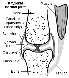
El lugar donde se encuentran dos huesos se llama articulación. Las articulaciones permiten el movimiento y la flexibilidad de varias partes del cuerpo. El movimiento de los huesos es causado por los músculos que tiran de los tendones que están unidos al hueso.
Cartilage covers the end of bones. Between the cartilage of two bones which form a joint there is a small amount of thick fluid called synovial fluid. This fluid lubricates the joint which allows smooth movement between the bones.
The synovial fluid is made by the tissue surrounding a joint (the synovium). The outer part of the synovium is called the capsule. This is tough, gives the joint stability and stops the bones from moving 'out of joint'. Surrounding ligaments and muscles also help to give support and stability to joints.
In the knee joint, the cartilage covering the lower part of the joint is thickened in the inner and outer part of the joint. These two areas of cartilage are sometimes called menisci. The menisci act like shock absorbers in the knee and are sometimes torn following a knee injury. Also, there are two strong cross-shaped (cruciate) ligaments in the middle of the knee joints, which are attached to the ends of the calf bone (tibia) and the thigh bone (femur). These also can be torn following a knee injury.
¿Existen posibles complicaciones?
Volver al contenidoIn most cases the procedure is done without any problems. Complications are generally rare but can include:
Accidental damage to structures inside or near to the joint.
Excessive bleeding inside the joint which can cause a lot of swelling and pain.
Infection within the joint - this can be serious.
As with any operation, there is a risk of allergy to local anaesthetics, or complications of anaesthesia if a general anaesthetic is used.
After arthroscopy or arthroscopic surgery, see a doctor urgently if you:
Have pain or swelling in the joint, which becomes worse. In particular, if the joint is also hot, tender and red. (This may indicate bleeding or infection in the joint.)
Develop a high temperature.
See fluid, pus or blood coming from the site of the cut (incision).
Develop numbness or tingling near to the joint (which may indicate nerve damage).
Selecciones del paciente para Otras cirugías y procedimientos

Cirugía y procedimientos
Reemplazo de rodilla
A knee replacement is an operation to replace damaged parts of the knee joint. It can be either a total knee replacement (TKR) or a partial (uni-compartmental) knee replacement. The new part of the joint is called a prosthesis.
por la Dra. Philippa Vincent, MRCGP

Cirugía y procedimientos
Abdominoplastia
In this procedure excess skin and fat can be removed, abdominal contours and scars improved, and the muscles tightened.
por la Dra. Jacqueline Payne, FRCGP
Lecturas adicionales y referencias
- Crawford R, Walley G, Bridgman S, et al; Imágenes por resonancia magnética frente a artroscopia en el diagnóstico de patologías de rodilla, concentrándose en lesiones meniscales y desgarros del LCA: una revisión sistemática. Br Med Bull. 2007;84:5-23. Epub 2007 Sep 3.
- Harris JD, Brophy RH, Siston RA, et al; Treatment of chondral defects in the athlete's knee. Arthroscopy. 2010 Jun;26(6):841-52.
Continúa leyendo abajo
Sobre el autorVer biografía completa

Dra. Rachel Hudson, MRCGP
Médico General y Autor Médico
MBChB, MRCGP (2008), BSc (Medical Science), DFSRH, DRCOG, DCH
La Dra. Rachel Hudson es una médica de cabecera del NHS que trabaja en el noroeste de Inglaterra.
Acerca del revisorVer biografía completa

Dra. Toni Hazell, MRCGP
MBBS, BSc, MRCGP, DFSRH, Dip GU med, DRCOG, DCH (London, UK, 2000)
La Dra. Toni Hazell se graduó de la Escuela de Medicina del Hospital St. Mary y realizó su VTS en el Hospital Northwick Park.
Historial del artículo
La información en esta página está escrita y revisada por pares por clínicos calificados.
Next review due: 19 Oct 2027
20 Oct 2024 | Última versión

Pregunta, comparte, conecta.
Navega por discusiones, haz preguntas y comparte experiencias en cientos de temas de salud.

¿Te sientes mal?
Evalúa tus síntomas en línea de forma gratuita
Suscríbete al boletín de Patient
Tu dosis semanal de consejos de salud claros y confiables, escritos para ayudarte a sentirte informado, seguro y en control.
Al suscribirte aceptas nuestros Política de Privacidad. Puedes darte de baja en cualquier momento. Nunca vendemos tus datos.