Escaneo de perfusión miocárdica
Revisado por pares por Dr Hayley Willacy, FRCGP Última actualización por Dr Colin Tidy, MRCGPÚltima actualización 20 Nov 2023
Cumple con las directrices editoriales
- DescargarDescargar
- Compartir
- Language
- Discusión
- Versión en audio
- Agregar a fuentes preferidas en Google
Un escaneo de perfusión miocárdica utiliza una pequeña cantidad de un químico radiactivo para ver qué tan bien fluye la sangre hacia los músculos del corazón (el miocardio). A menudo, este escaneo se realiza después de un ejercicio suave para ver cómo responde el músculo cardíaco bajo estrés.
De un vistazo
Myocardial perfusion scans help find the cause of unexplained chest pain.
This scan shows blood flow patterns to the heart walls.
A small amount of a radioactive chemical is injected, which concentrates in the heart muscle.
A special camera detects gamma rays from the chemical to create an image of your heart.
The scan can be done while resting or after gentle exercise.
Tell your doctor if you are pregnant or breastfeeding, as special precautions may be needed.
Side effects are rare but can include abnormal heart rhythm or wheezing.
What is the myocardium?
The heart is mainly made of special muscle called the myocardium. The muscle pumps blood into arteries (blood vessels) which take the blood to every part of the body.
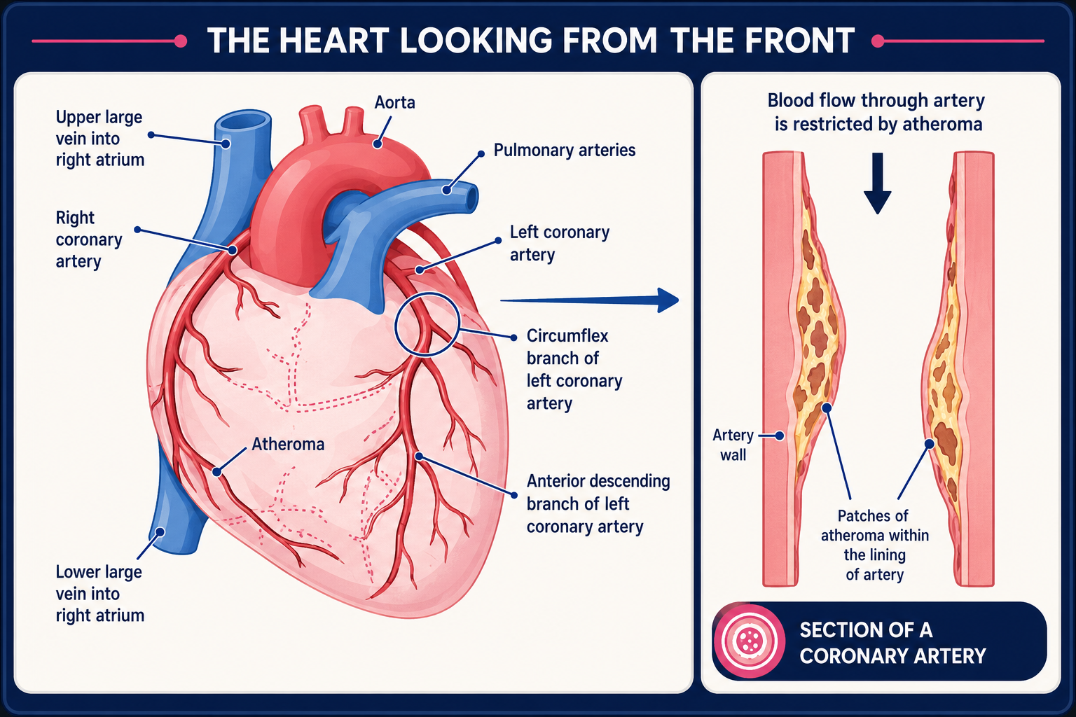
Like any other muscle, the myocardium needs a good blood supply. When the blood supply to the heart is reduced it may 'complain' with pain; this pain is called angina. The heart (coronary) arteries supply the heart with blood.
The usual cause of angina is narrowing of one or more of the coronary arteries. The blood supply may be enough when resting. However, the heart muscle needs more blood and oxygen when it works harder.
For example, when walking fast or climbing stairs, the heart rate increases to deliver the extra blood. If the extra blood that the heart needs during exertion cannot get past the narrowed arteries, the heart 'complains' with angina pain. The diagram below illustrates how angina occurs.
Corazón con ateroma
What is a myocardial perfusion scan used for?
Myocardial perfusion scans can be used to try to find the cause of unexplained chest pain, or chest pain brought on by exercise. This test may also be done to:
Show blood flow patterns to the heart walls.
See whether the heart (coronary) arteries are blocked and by how much.
Determine the extent of injury to the heart following a heart attack (myocardial infarction).
How does a myocardial perfusion scan work?
A myocardial perfusion scan uses a special chemical called a radionuclide. A radionuclide (sometimes called a radioisotope or isotope) is a chemical which sends out (emits) a type of radioactivity called gamma rays. In a myocardial perfusion scan a tiny amount of radionuclide is put into the body, usually by an injection into a vein.
Existen diferentes tipos de radionúclidos. Algunos tienden a acumularse o concentrarse en diferentes órganos o tejidos. Por lo tanto, el radionúclido utilizado depende de qué parte del cuerpo se va a escanear.
A radionuclide that concentrates in heart muscle is used for a myocardial perfusion scan. Other scans that use different radionuclides include a bone scan and a thyroid scan. See separate leaflets called Escaneo Óseo y Thyroid Scans and Uptake Tests para más detalles.
The radionuclide travels through the bloodstream and into the heart muscle. As the radionuclide moves through the heart muscle, areas that have good blood flow take up (absorb) the radionuclide well.
Areas that do not absorb radionuclide very well may have a poor blood supply due to narrowed heart (coronary) arteries, or may have been damaged by a heart attack. So, heart muscle tissue with a good blood flow will emit more gamma rays than areas with a poor blood flow or damaged tissue.
Los rayos gamma son similares a los rayos X y son detectados por un dispositivo llamado cámara gamma. Los rayos gamma que se emiten desde el interior del cuerpo son detectados por la cámara gamma, se convierten en una señal eléctrica y se envían a una computadora.
The computer builds a picture by converting the differing intensities of radioactivity emitted into different colours or shades of grey.
For example, areas of the target organ or tissue (in this case, the heart) which emit lots of gamma rays may be shown as red spots ('hot spots') on the picture on the computer monitor.
Las áreas que emiten bajos niveles de rayos gamma pueden mostrarse en azul ('puntos fríos'). Se pueden usar varios otros colores para los niveles 'intermedios' de rayos gamma emitidos.
This creates a picture that shows which parts of the heart muscle have good blood flow and which parts do not.
What happens during a myocardial perfusion scan?
The myocardial perfusion scans can be carried out in a number of ways. The test can be carried out while you are resting, after gentle exercise, or on two separate occasions to combine resting and exercising. The exact order in which the test is carried out varies between different hospitals.
Exercise test
In an exercise test you may be given an injection of a medication that makes your heart beat faster and stronger. Some people find that this gives them a tingling feeling in the chest or a sensation that their heart is beating strongly (palpitations).
These sensations usually pass quickly after the test is over. When the heart is beating at a certain rate the radionuclide chemical is injected, usually into the hand.
If you are able to, you may be asked to do some pedalling on an exercise bicycle during your test. The amount of activity you are asked to do will depend on your individual condition.
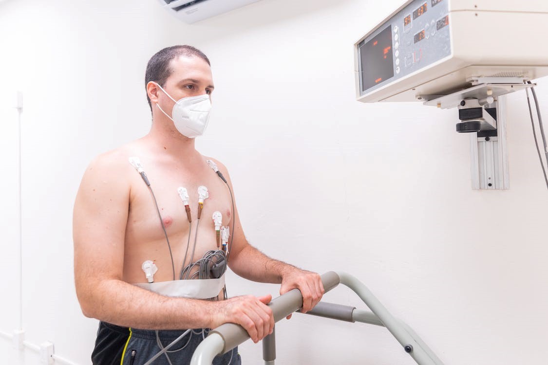
You will usually be monitored during the test with a heart trace called an electrocardiogram (ECG). This means that several sticky pads, called electrodes, will be placed on your chest. The electrodes are connected to a machine which shows how your heart responds.
Sometimes you will have a set of images taken soon after the radionuclide chemical is given. Some hospitals ask you to eat something before the pictures are taken. This can help to make the images clearer.
The scan itself
When it is time to do the scanning, you lie on a couch while the gamma camera detects the gamma rays coming from your body. The computer turns the information into a picture. You need to lie as still as possible whilst each picture is taken (so it is not blurred). Actual scanning time for each heart scan varies from 16-30 minutes, depending on the type of scanner used.
Depending on the reason for your test you may need to have a second scan. This may take place on the same day, 24 hours later or a few days later. You will usually only have the exercise test once. So, for the second test you will just receive an injection of the radionuclide chemical and then have the images taken.
Resting test
A resting test is carried out in the same way, except that there is no injection of a medication to make your heart beat faster and there is no exercise.
What should I do to prepare
Your local hospital should give you specific information to help you prepare for these tests. As these tests involve a small amount of radiation, pregnant women should not have them. Let your doctor know if you are, or think you could be, pregnant. You should also let your doctor know if you are breast-feeding.
Generally there is not much preparation needed before this test. However, you may be asked not to eat or drink anything that contains caffeine before the test. In some cases your doctor may advise you not to take your medication for a few days before the scan.
You may also be asked to bring a list of medication along with you on the day of the test. Your local hospital will advise if this applies to you.
What can I expect after a myocardial perfusion scan?
Myocardial perfusion scans do not generally cause any after effects. Through the natural process of radioactive decay, the small amount of radioactive chemical in your body will lose its radioactivity over time.
It may also pass out of your body through your urine or stool (faeces) during the first few hours or days following the test. You may be instructed to take special precautions after urinating, to flush the toilet twice and to wash your hands thoroughly.
If you have contact with children or pregnant women you should let your doctor know. Although the levels of radiation used in the scan are small, your doctor may advise special precautions. Your local hospital should give you more advice on this.
Are there any side-effects or complications?
Most people have a myocardial perfusion scan without any problems. It is possible, although rare, that the exercise or the medication that makes the heart beat faster could cause an abnormal heart rhythm (arrhythmia) or heart attack (myocardial infarction).
The medication that makes the heart beat faster may, occasionally, make some people 'wheezy'. The risk of this happening is higher if you have asthma or other lung conditions.
The term 'radioactivity' may sound alarming. However, the levels of radioactive chemicals used in radionuclide scans are very small and considered to be safe, and they leave the body quickly. The dose of radiation that the body receives is very small. In many cases, the level of radiation involved is not much different to a series of a few normal X-rays. However:
As with any other types of radiation (such as X-ray), there is a small risk that the gamma rays may affect an unborn child. So, tell your doctor if you are pregnant or if you may be pregnant.
Raramente, algunas personas tienen una reacción alérgica al químico inyectado.
Teóricamente, es posible recibir una sobredosis cuando se inyecta el químico. Esto es muy raro.
Selecciones del paciente para Pruebas cardíacas

Pruebas e investigaciones
Cateterismo cardíaco
La cateterización cardíaca es una forma de obtener información detallada sobre su corazón y las arterias coronarias, y también es posible ofrecer tratamiento para algunas condiciones al mismo tiempo.
por el Dr. Colin Tidy, MRCGP
Pruebas e investigaciones
Prueba de esfuerzo
Una prueba de tolerancia al ejercicio (ETT) registra la actividad eléctrica del corazón durante el ejercicio. Es más útil en pacientes que experimentan dolor en el pecho al hacer esfuerzo. También se utiliza para detectar si las anomalías en el ritmo cardíaco pueden ser provocadas por el ejercicio.
por la Dra. Philippa Vincent, MRCGP
Preguntas frecuentes
What is the specific radionuclide used in a myocardial perfusion scan?
The article states that a radionuclide that concentrates in heart muscle is used for a myocardial perfusion scan. However, it does not specify the exact chemical name of this radionuclide.
How long does the radionuclide stay in the body after the scan?
The radioactive chemical used in the scan loses its radioactivity over time through natural decay. It also leaves the body through urine or stool (faeces) during the first few hours or days following the test. You might be asked to take special precautions, like flushing the toilet twice after urinating and washing your hands thoroughly.
Are there different types of equipment used for myocardial perfusion scans?
The article mentions that the actual scanning time for each heart scan varies from 16-30 minutes, depending on the type of scanner used. This implies that there are different types of gamma cameras or scanning equipment that might be utilised for the procedure.
What kind of specific dietary restrictions, besides caffeine, might I have before the scan?
The article primarily mentions that you may be asked not to eat or drink anything containing caffeine before the test. It does not elaborate on other specific dietary restrictions, but advises that your local hospital will give you specific preparation information.
How long will the entire myocardial perfusion scan appointment take, including preparation and recovery time?
The article details the various steps involved, such as potential exercise tests, injection, and scanning times ranging from 16-30 minutes per scan. It also mentions that sometimes a second scan is needed on the same day, 24 hours later, or a few days later. However, it does not provide an overall estimate for the total duration of the appointment or the entire process from start to finish.
Lecturas adicionales y referencias
- Gammagrafía de perfusión miocárdica para el diagnóstico y manejo de la angina e infarto de miocardio; Guía de Evaluación de Tecnología de NICE, noviembre de 2003 (última actualización julio de 2011)
- Sakatani T, Nakajima K, Nishimura T; Cardiovascular event risk estimated by myocardial perfusion SPECT combined with clinical data. J Cardiol. 2022 Jul;80(1):64-71. doi: 10.1016/j.jjcc.2021.10.004. Epub 2021 Oct 30.
Sobre el autorVer biografía completa

Dr Colin Tidy, MRCGP
Médico General, Autor Médico
MBBS, MRCGP, MRCP (Paediatrics), DCH
El Dr. Colin Tidy es un médico del NHS, con sede en Oxfordshire.
Acerca del revisorVer biografía completa

Dr Hayley Willacy, FRCGP
Médico General, Autor Médico
MBChB (1992), DRCOG, DFFP, MRCOG (Part 1) MRCGP (2007), DFSRH (2013), MSc - medical education (2020)
La Dra. Hayley Willacy fue una médica general del NHS que trabajaba en el noroeste de Inglaterra, quien se retiró de la práctica clínica en 2022 después de 30 años.
Historial del artículo
La información en esta página está escrita y revisada por pares por clínicos calificados.
Artículo también disponible en Inglés, Alemán, Español, Francés, Italiano, Portugués, Hindi, Hebreo, Árabe, y Sueco.
Próxima revisión: 18 Nov 2028
20 Nov 2023 | Última versión

Pregunta, comparte, conecta.
Navega por discusiones, haz preguntas y comparte experiencias en cientos de temas de salud.

¿Te sientes mal?
Evalúa tus síntomas en línea de forma gratuita
Suscríbete al boletín de Patient
Tu dosis semanal de consejos de salud claros y confiables, escritos para ayudarte a sentirte informado, seguro y en control.
Al suscribirte aceptas nuestros Política de Privacidad. Puedes darte de baja en cualquier momento. Nunca vendemos tus datos.
Más en pruebas e investigaciones
- Grupos y tipos sanguíneos
- Genes BRCA
- Detección de cáncer de mama
- Broncoscopia
- Angiografía cerebral
- Cistoscopia
- Electrocardiograma
- Prueba de esfuerzo
- Prueba inmunoquímica fecal
- Aspiración con aguja fina
- Hiponatremia
- prueba del talón del recién nacido
- prueba de detección en recién nacidos
- Programa de cribado del NHS
- pruebas de detección durante el embarazo
- Proteinuria
- Sigmoidoscopia
- Espirometría
- Pruebas de ITS
- Talasemia