Nevus sebáceo
Revisado por pares por Dr Philippa Vincent, MRCGPÚltima actualización por Dr Doug McKechnie, MRCGPLast updated 24 de mayo de 2023
Cumple con las directrices editoriales
- DescargarDescargar
- Compartir
- Language
- Discusión
- Versión en audio
- Add to preferred sources on Google
Profesionales Médicos
Professional Reference articles are designed for health professionals to use. They are written by UK doctors and based on research evidence, UK and European Guidelines. You may find one of our artículos de salud more useful.
En este artículo:
Sinónimos: nevus sebáceo, nevus sebáceo de Jadassohn, nevus epidérmico verrugoso, nevus organoide. Nota la ortografía americana 'nevus'.
Continúa leyendo abajo
¿Qué es un nevus sebáceo?
A nevus sebáceo is a congenital skin lesion which usually manifests as a bald patch on the scalp of the baby. The area of hair loss looks orange or yellowish, with a velvety feel and appearance. It is usually oval or circular. In adolescence, under the influence of sex hormones, it becomes bumpy and warty, with an unpleasant verrucoid appearance.
Durante la adolescencia y la adultez temprana, generalmente permanecen inactivos. A veces se agrandan en la adultez tardía y, raramente, sufren un cambio neoplásico que en general implica tumores benignos.
Los nevos sebáceos son hamartomas, lo que significa que se originan a partir de tejidos que normalmente se encuentran en ese sitio, pero crecen de manera desorganizada. Tienen un origen embriológico común de la unidad pilosebácea-apocrina y, por lo tanto, contienen glándulas sebáceas, folículos pilosos y glándulas sudoríparas. Por lo tanto, el término nevo sebáceo es un poco inexacto: algunos patólogos prefieren el término nevo epidérmico verrugoso o nevo organoide.1 Although they contain elements of hair follicles, normal terminal hair follicles are absent making the area look hairless. Most are solitary, with the scalp being the most common site.
Nevus sebáceo del cuero cabelludo
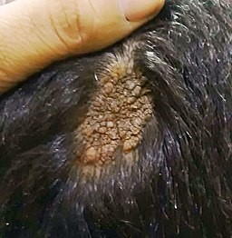
© Mohammad2018, CC BY-SA 4.0, a través de Wikimedia Commons
Nevus sebáceo (Epidemiología)
Volver al contenidoLa prevalencia estimada es del 0.3% de todos los recién nacidos en todo el mundo, con una distribución equitativa entre sexos.2
Continúa leyendo abajo
Síntomas del nevus sebáceo (presentación)
Volver al contenidoPor lo general, se observa un solo parche sin pelo (redondo o lineal) en el cuero cabelludo al nacer o poco después.
Se puede observar la apariencia clásica aterciopelada de color tostado o amarillo-naranja.
La lesión puede parecer más prominente en los recién nacidos, debido a los efectos de las hormonas sexuales maternas.
En los adolescentes, la lesión tiende a adoptar su apariencia nodular verrucosa debido a la influencia de las hormonas sexuales.
En la vida posterior, puede haber un crecimiento excesivo de componentes de la lesión que indique el posible desarrollo de tumores benignos o malignos dentro de la lesión.
Diagnóstico diferencial
Volver al contenidoLa apariencia de los nevos sebáceos es bastante característica, pero las siguientes lesiones pueden tener una apariencia similar o afectar áreas similares del cuerpo:
Aplasia cutis congénita.
Naevus syringocystadenomatosus papilliferous.
Xantogranuloma juvenil.
Mastocitoma solitario.
Other causes of alopecia.
Continúa leyendo abajo
Investigaciones
Volver al contenidoLa biopsia puede considerarse con fines diagnósticos o para investigar la naturaleza de los tumores que surgen dentro de ellos.
Enfermedades asociadas
Volver al contenidoAproximadamente un tercio de las personas con un nevus sebáceo tienen anomalías sistémicas que afectan al cerebro, los ojos o los sistemas esqueléticos. Cuando estas ocurren, se denomina síndrome del nevus sebáceo.3 In these cases the naevi follow the lines of Blaschko; the more extensive the cutaneous involvement, the greater the risk of extracutaneous manifestations.4 Subsequent problems can include infantile epilepsy, developmental delay and intellectual disability.
Tratamiento y manejo del nevo sebáceo5
Volver al contenidoDebido a la dificultad de cuantificar el riesgo de transformación maligna, algunos especialistas abogan por la excisión de piel de espesor completo antes de la adolescencia.6 Others recommend watchful waiting.5
En algunos casos, de todos modos, no serán adecuados para la extirpación debido a su tamaño y ubicación. Estos deben someterse a revisiones regulares.
La terapia fotodinámica puede desempeñar un papel en la eliminación de la lesión.7
Consulte el consejo de un dermatólogo y/o cirujano plástico para ayudar a decidir sobre la necesidad y el momento de la excisión.
Cualquier lesión que cambie significativamente su apariencia, se ulcere, se vuelva dolorosa, sangre o muestre otros signos de posible malignidad, debe ser remitida a un dermatólogo.
Complicaciones
Volver al contenidoApariencia cosmética deficiente.
Desarrollo de un tumor benigno o maligno dentro de la lesión.
Complicaciones asociadas con la extirpación de la lesión.
Pronóstico
Volver al contenidoLa mayoría de las lesiones permanecen inactivas durante toda la vida adulta, siendo la única preocupación de tipo cosmético. Dos series de casos que estudiaron casi 1,300 nevus sebáceos encontraron que el 15% experimentan cambios neoplásicos.8 9
De este 15%, el 80% desarrolla focos de neoplasias benignas como tricoblastomas y siringocistadenomas. El 20% restante desarrolla cambios malignos, con carcinomas basocelulares que constituyen la mitad de estos, y carcinomas de células escamosas un cuarto.
Por lo general, cualquier cambio maligno ocurre en la edad adulta, pero se ha reportado en niños de tan solo 8 años.6 Malignancy can be diagnosed in the same way as other malignant skin tumours, with dermoscopy playing its usual role.10
Exclusive updates for healthcare professionals
Stay informed with the latest clinical updates, professional insights, and evidence-based guidance. The Patient Pro newsletter curates essential content for healthcare professionals—delivered straight to your inbox.
By subscribing you accept our Política de Privacidad. Puedes darte de baja en cualquier momento. Nunca vendemos tus datos.
Lecturas adicionales y referencias
- Simi CM, Rajalakshmi T, Correa M; Análisis clinicopatológico de 21 casos de nevus sebáceo: un estudio retrospectivo. Indian J Dermatol Venereol Leprol. 2008 Nov-Dic;74(6):625-7.
- Moody MN, Landau JM, Goldberg LH; Nevus sebáceo revisitado. Pediatr Dermatol. 2012 Ene-Feb;29(1):15-23. doi: 10.1111/j.1525-1470.2011.01562.x. Publicado electrónicamente 2011 Oct 13.
- Laura FS; Síndrome del nevus epidérmico. Handb Clin Neurol. 2013;111:349-68. doi: 10.1016/B978-0-444-52891-9.00041-5.
- Asch S, Sugarman JL; Síndromes de nevus epidérmico. Handb Clin Neurol. 2015;132:291-316. doi: 10.1016/B978-0-444-62702-5.00022-6.
- Nevus sebáceo; DermNet NZ
- Rosen H, Schmidt B, Lam HP, et al; Manejo del nevus sebáceo y el riesgo de carcinoma de células basales: una revisión de 18 años. Pediatr Dermatol. 2009 Nov-Dic;26(6):676-81. doi: 10.1111/j.1525-1470.2009.00939.x. Publicado en línea el 20 de julio de 2009.
- Babilas P, Szeimies RM; El uso de la terapia fotodinámica en dermatología. G Ital Dermatol Venereol. 2010 Oct;145(5):613-30.
- Idriss MH, Elston DM; Neoplasias secundarias asociadas con nevus sebáceo de Jadassohn: un estudio de 707 casos. J Am Acad Dermatol. 2014 Feb;70(2):332-7. doi: 10.1016/j.jaad.2013.10.004. Epub 20 de noviembre de 2013.
- Cribier B, Scrivener Y, Grosshans E; Tumores que surgen en nevus sebáceo: Un estudio de 596 casos. J Am Acad Dermatol. 2000 Feb;42(2 Pt 1):263-8.
- Enei ML, Paschoal FM, Valdes G, et al; Carcinoma de células basales que aparece en un nevus sebáceo facial de Jadassohn: características dermoscópicas. An Bras Dermatol. 2012 Jul-Aug;87(4):640-2.
Continúa leyendo abajo
About the authorView full bio

Dr Doug McKechnie, MRCGP
Medical Writer
MA, MBBS, MSc, DRCOG, MRCP(UK), MRCGP(2021), FHEA
Dr Doug McKechnie is an NHS GP working in London. He works full-time clinically and is also the Deputy Lead for the Clinical and Professional Practice module at University College London Medical School.
About the reviewerView full bio

Dra. Philippa Vincent, MRCGP
Médico General, Autor Médico
MB BS, Bsc, MRCGP (2000), DCH, DFSRH, DRCOG
Dra Philippa Vincent is an NHS GP working in North London.
Historial del artículo
La información en esta página está escrita y revisada por pares por clínicos calificados.
Próxima revisión: 12 de mayo de 2028
24 de mayo de 2023 | Última versión

Pregunta, comparte, conecta.
Navega por discusiones, haz preguntas y comparte experiencias en cientos de temas de salud.

¿Te sientes mal?
Evalúa tus síntomas en línea de forma gratuita