Optic atrophy type 1
Última actualización 8 Nov 2023
Cumple con las directrices editoriales
- DescargarDescargar
- Compartir
- Language
- Discusión
- Versión en audio
- Agregar a fuentes preferidas en Google
Optic atrophy type 1 is a condition that often causes slowly worsening vision, usually beginning in childhood. People with optic atrophy type 1 typically experience a narrowing of their field of vision (tunnel vision). Affected individuals gradually lose their sight as their field of vision becomes smaller. Both eyes are usually affected equally, but the severity of the vision loss varies widely, even among affected members of the same family, ranging from nearly normal vision to complete blindness.
Descripción
Optic atrophy type 1 is a condition that often causes slowly worsening vision, usually beginning in childhood. People with optic atrophy type 1 typically experience a narrowing of their field of vision (tunnel vision). Affected individuals gradually lose their sight as their field of vision becomes smaller. Both eyes are usually affected equally, but the severity of the vision loss varies widely, even among affected members of the same family, ranging from nearly normal vision to complete blindness.
In addition to vision loss, people with optic atrophy type 1 frequently have problems with colour vision (colour vision deficiency) that make it difficult or impossible to distinguish between shades of blue and green.
In the early stages of the condition, individuals with optic atrophy type 1 experience a progressive loss of certain cells within the retina, which is a specialized light-sensitive tissue that lines the back of the eye. The loss of these cells (known as retinal ganglion cells) is followed by the degeneration (atrophy) of the nerves that relay visual information from the eye to the brain (optic nerves), which results in further vision loss. Atrophy causes these nerves to have an abnormally pale appearance (pallor), which can be seen during an eye examination.
Patrones de herencia
The source lists the following possible inheritance patterns: Autosomal dominant. More than one pattern can apply in different cases.
Related genes
Otros nombres
ADOA
Autosomal dominant optic atrophy
Autosomal dominant optic atrophy Kjer type
DOA
Dominant optic atrophy
Kjer type optic atrophy
Kjer's optic atrophy
Optic atrophy, autosomal dominant
Optic atrophy, hereditary, autosomal dominant
Optic atrophy, juvenile
Optic atrophy, Kjer type
Related patient information and medicines

Patient information leaflet
Problemas oculares
This leaflet contains a list of conditions which affect the eye, grouped together by the part of the eye which is affected or involved. See the leaflet called Anatomy of the eye for details on the structure of the eye. See the separate leaflets called Diabetic retinopathy and High blood pressure (Hyper. Optic atrophy Optic atrophy is the loss of.

Patient information leaflet
Diabetes
There are two main types of diabetes - diabetes tipo 1 and type 2 diabetes. Type 1 usually first develops in children or in young adults. This leaflet refers to diabetes mellitus (now usually just known as diabetes); there is a separate condition llamado diabetes insípida which is always called by its full name. It is also called DIDMOAD (because.

Patient information leaflet
Diabetes tipo 1
Diabetes tipo 1 es the type of diabetes that typically develops in children and in young adults. In type 1 diabetes the body stops making insulin and the blood sugar (glucose) level... Treatment of complications Too much insulin can make the blood glucose level go too low ( hipoglucemia , sometimes called a 'hypo'). See the separate leaflets.

Patient information leaflet
Problemas visuales
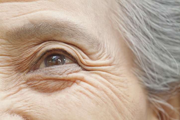
Visual problems can arise from problems affecting different parts of the eye. Generally speaking, the further towards the back of the eye that problems occur, the more likely they are to have a long-lasting or permanent effect on your eyesight. Retinal dystrophy Retinal dystrophies son a group of inherited (genetic) disorders which result in.
Patient information leaflet
Degeneración macular
Degeneración macular relacionada con la edad is the most common cause of sight impairment in those aged over 50. It causes a gradual loss of central vision, which is needed for detailed work and for things like reading and driving. Macular degeneration (also known as age-related macular degeneration) occurs when cells in the mácula - the central area of the.

Patient information leaflet
Fumar
Cigarette smoking is the greatest single cause of illness and premature death en the UK. Worldwide, tobacco kills 7 million people every year. Almost a million deaths are due to non-smokers being exposed to second-hand smoke. An overactive thyroid gland (hyp... A disorder of the brain and spinal cord ( multiple sclerosis ). Inflammation of the.
Lecturas adicionales y referencias
- Optic atrophy type 1; MedlinePlus, National Library of Medicine.
- Delettre-Cribaillet C, Hamel CP, Lenaers G. Optic Atrophy Type 1 - RETIRED CHAPTER, FOR HISTORICAL REFERENCE ONLY. 2007 Jul 13 [updated 2015 Nov 12]. In: Adam MP, Bick S, Mirzaa GM, Wallace SE, Amemiya A, editors. GeneReviews(R) [Internet]. Seattle (WA): University of Washington, Seattle; 1993-2026. Available from http://www.ncbi.nlm.nih.gov/books/NBK1248/
- Ferre M, Bonneau D, Milea D, Chevrollier A, Verny C, Dollfus H, Ayuso C, Defoort S, Vignal C, Zanlonghi X, Charlin JF, Kaplan J, Odent S, Hamel CP, Procaccio V, Reynier P, Amati-Bonneau P. Molecular screening of 980 cases of suspected hereditary optic neuropathy with a report on 77 novel OPA1 mutations. Hum Mutat. 2009 Jul;30(7):E692-705. doi: 10.1002/humu.21025.
- Formichi P, Radi E, Giorgi E, Gallus GN, Brunetti J, Battisti C, Rufa A, Dotti MT, Franceschini R, Bracci L, Federico A. Analysis of opa1 isoforms expression and apoptosis regulation in autosomal dominant optic atrophy (ADOA) patients with mutations in the opa1 gene. J Neurol Sci. 2015 Apr 15;351(1-2):99-108. doi: 10.1016/j.jns.2015.02.047. Epub 2015 Mar 6.
- Roubertie A, Leboucq N, Picot MC, Nogue E, Brunel H, Le Bars E, Manes G, Angebault Prouteau C, Blanchet C, Mondain M, Chevassus H, Amati-Bonneau P, Sarzi E, Pages M, Villain M, Meunier I, Lenaers G, Hamel CP. Neuroradiological findings expand the phenotype of OPA1-related mitochondrial dysfunction. J Neurol Sci. 2015 Feb 15;349(1-2):154-60. doi: 10.1016/j.jns.2015.01.008. Epub 2015 Jan 13.
- Yu-Wai-Man P, Griffiths PG, Burke A, Sellar PW, Clarke MP, Gnanaraj L, Ah-Kine D, Hudson G, Czermin B, Taylor RW, Horvath R, Chinnery PF. The prevalence and natural history of dominant optic atrophy due to OPA1 mutations. Ophthalmology. 2010 Aug;117(8):1538-46, 1546.e1. doi: 10.1016/j.ophtha.2009.12.038. Epub 2010 Apr 24.
- Zanna C, Ghelli A, Porcelli AM, Karbowski M, Youle RJ, Schimpf S, Wissinger B, Pinti M, Cossarizza A, Vidoni S, Valentino ML, Rugolo M, Carelli V. OPA1 mutations associated with dominant optic atrophy impair oxidative phosphorylation and mitochondrial fusion. Brain. 2008 Feb;131(Pt 2):352-67. doi: 10.1093/brain/awm335.
Únete a la conversación
Comparte tus pensamientos o experiencias con otros en nuestro comunidades de pacientes.

Pregunta, comparte, conecta.
Navega por discusiones, haz preguntas y comparte experiencias en cientos de temas de salud.
Suscríbete al boletín de Patient
Tu dosis semanal de consejos de salud claros y confiables, escritos para ayudarte a sentirte informado, seguro y en control.
Al suscribirte aceptas nuestros Política de Privacidad. Puedes darte de baja en cualquier momento. Nunca vendemos tus datos.