Pleural effusion
Peer reviewed by Dr Doug McKechnie, MRCGPLast updated by Dr Hayley Willacy, FRCGP Last updated 21 Dec 2023
Meets Patient’s editorial guidelines
- DownloadDownload
- Share
- Language
- Discussion
- Audio Version
- Add to preferred sources on Google
A pleural effusion is a collection of fluid next to the lung. There are various causes. The effusion may cause you to become breathless. The fluid can be drained if necessary. Treatment is mainly aimed at the underlying cause.
At a glance
A pleural effusion is a build-up of fluid in the space between a lung and the chest wall.
This condition can be caused by various factors, including infections, heart failure, and some cancers.
Symptoms can include chest pain and breathlessness, especially as the fluid increases.
A chest X-ray typically confirms a pleural effusion.
Treatment often focuses on the underlying cause, but the fluid itself can also be drained.
Procedures like pleurodesis or permanent drains can help manage recurrent effusions.
What is a pleural effusion?
Lungs and airways with pleura
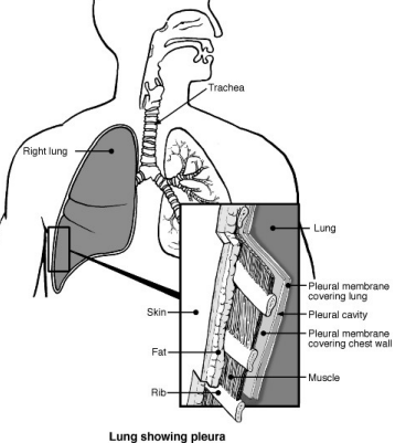
A pleural effusion means that there is a build-up of fluid in the space between a lung and the chest wall.
The pleura is a thin membrane that lines the inside of the chest wall and covers the lungs. There is normally a tiny amount of fluid between the two layers of pleura. This acts like lubricating oil between the lungs and the chest wall as they move when you breathe.
A pleural effusion develops when this fluid builds up and separates the lung from the chest wall.
Types of pleural effusions
Pleural effusions are classed as transudates or exudates according to how much protein they contain.
Transudates have a low protein level of <25g/L. Fluid accumulates due to a disruption in the balance of fluid pressures.
Exudates have a high protein level of >35g/L. Fluid accumulates due to increased leakiness (permeability) of the smallest blood vessels.
Transudative pleural effusions
These are most often caused by conditions such as heart failure and liver failure. Less common causes are nephrotic syndrome, hypothyroidism and as a consequence of peritoneal dialysis.
Exudative pleural effusions
The most common causes of these are infections, such as pneumonia or tuberculosis, or cancer. Less common causes include autoimmune conditions such as rheumatoid arthritis, after a heart attack and pancreatitis.
Causes of a pleural effusion
A pleural effusion is a complication of various conditions. The following are some of the more common causes of a pleural effusion (but there are other rarer causes too):
Lung infection (pneumonia), tuberculosis, and cancers may cause inflammation of the lung and pleura. This may cause fluid to build up into a pleural effusion.
Some arthritic conditions may cause inflammation of the pleura in addition to joint inflammation. For example, pleural effusion is an uncommon complication of rheumatoid arthritis and systemic lupus erythematosus (SLE).
Heart failure causes 'back pressure' in the veins (blood vessels) that take blood back to the heart. Some fluid may seep out of the blood vessels. Swelling of the legs with fluid is typical with heart failure, but a pleural effusion may also develop.
A low level of protein in the blood also tends to allow fluid to seep out of the blood vessels. For example, cirrhosis of the liver and some kidney diseases may cause a low level of blood protein which allows a pleural effusion to develop.
Symptoms of a pleural effusion
You may feel some chest pain but a pleural effusion is often painless. The amount of fluid varies. As the effusion becomes larger, it presses on the lung, which cannot expand fully when you breathe. You may then become breathless.
You may also have symptoms of the condition that is causing the effusion. As a whole range of conditions can cause a pleural effusion, there is a large range of other symptoms that may occur, depending on the underlying cause. One example is you may have a cough and a high temperature (fever) if the cause is lung infection (pneumonia).
How is a pleural effusion diagnosed?
A chest X-ray usually confirms a build-up of fluid between a lung and the chest wall (pleural effusion). If the cause of the effusion is known then no further tests may be needed. However, sometimes a pleural effusion is the first sign of an underlying condition.
Further tests may then be advised to find the cause of the effusion. These may include lung tests, blood tests and taking a sample of the fluid and pleura to examine in the laboratory.
Pleural effusion treatment
Treating the underlying cause
A major part of treatment is usually directed to the underlying cause of the build-up of fluid between the lung and the chest wall (pleural effusion). For example, medicines called antibiotics for lung infection (pneumonia), chemotherapy or radiotherapy for cancers, etc.
Therefore, treatment can vary greatly, depending on the cause of the effusion. If the underlying cause can be successfully treated then there is a good chance that the pleural effusion will go away for good. If the underlying cause cannot be treated, or can only be partially treated, the effusion may return if it is cleared (drained).
Treating the effusion itself
Small effusions that cause no symptoms, or only mild symptoms, may just be left and 'observed'. Treatment is usually only needed if the effusion causes symptoms such as breathlessness.
A large pleural effusion that makes you breathless can be drained. This is called a pleural fluid aspiration or pleural tap. It is usually done by inserting a needle or tube through the chest wall. A local anaesthetic is injected into the skin and chest wall first to make the procedure painless. This may be a 'one-off' procedure to relieve symptoms.
Lungs and airways - pleural effusion
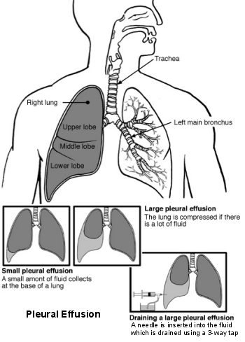
However, in many cases, unless the underlying cause can be treated, an effusion is likely to return within a few weeks. Repeated draining of the fluid, when symptoms become troublesome, is one option.
Depending on the underlying cause, other treatment options that are sometimes considered include:
Pleurodesis.
In this procedure, a special chemical (a sclerosant) is injected into the pleural space. This causes inflammation of the pleural membranes and helps them to 'stick' together. This helps to prevent fluid building up again into an effusion. Pleurodesis is most often used in the treatment of repeated (recurrent) effusions caused by cancer.
Talc pleurodesis is often used. If the lung re-inflates after the fluid has been drained, sterile talcum powder (talc) can be used to help stick the pleura together. The doctor puts the talc through the tube attached to the drain and then leaves the drain clamped for about an hour. This allows time for the 2 linings of the lung to stick together. The doctor may attach the drain to a suction machine to apply a small amount of pressure. This can help the pleura to seal together. After a pleurodesis, you will usually have the drain in place for another 24 hours.
Permanent drains
If the effusion is recurrent (perhaps because the underlying cause is not treatable), it is possible to have repeated chest drains put in. This may be uncomfortable and mean spending a lot of time going in and out of the hospital. In this situation, a special catheter called a tunnelled indwelling pleural catheter (TIPC), can be put in. This allows the pleural effusion to be drained easily while you are at home. When you feel you have a build-up of fluid on the lung, the catheter allows the fluid to be drained off into a bottle. This can be done by you, a family member or a nurse. The catheter is inserted in hospital on a day ward. As long as there are no complications, you can usually go home on the same day.
Shunt insertion
An operation to insert a shunt (like an internal drain) to allow the fluid to drain out from the chest into the tummy (abdominal) cavity. This is called a 'pleuroperitoneal shunt'. It is only occasionally used.
Video assisted thorascopic surgery
It may be possible to drain a pleural effusion and do a pleurodesis using a procedure called a thoracoscopy. This is a type of keyhole surgery where the doctor puts a small flexible tube (a thorascope) into the chest. The tube has a light and camera at the end, so the doctors can see into your chest. This is why it is called 'video assisted'. It can be used for taking a biopsy, for draining an effusion or pleurodesis. For this procedure, you lie on your side and are given an injection of a sedative to make you feel drowsy. Local anaesthetic is used to numb the area and the doctor makes a small cut to put the thorascope in. The procedure takes about 40 to 60 minutes and afterwards a chest drain will be left to drain any remaining fluid from the chest cavity.
Surgery also includes an operation to remove the pleura called a pleurectomy. It is sometimes used in people with effusions due to cancer when other treatment options have failed.
Patient picks for Lung conditions
Chest and lungs
Carbon monoxide poisoning
Carbon monoxide is an odourless poisonous gas. Even small amounts can deprive the body of oxygen, and can lead to brain damage in severe cases. This leaflet describes the symptoms of carbon monoxide and advises on how to protect yourself from carbon monoxide poisoning.
by Dr Philippa Vincent, MRCGP
Chest and lungs
Pulmonary fibrosis
Pulmonary fibrosis is a serious lung disease where the tiny air sacs of the lungs (the alveoli) and the lung tissue next to the alveoli become damaged and scarred, resulting in lung fibrosis. The main symptom is shortness of breath that gradually gets worse. The cause is often not known but pulmonary fibrosis can be caused by a variety of underlying conditions. Treatments include treating any underlying cause in addition to steroids and other medicines. Increasingly lung transplantation may be considered.
by Dr Toni Hazell, FRCGP
Frequently asked questions
How long can a pleural effusion last if left untreated?
The duration of a pleural effusion varies significantly depending on its underlying cause. Small effusions that cause no symptoms may be left and observed, potentially resolving on their own. However, if the underlying condition is not treated, larger effusions are likely to return or persist.
What is the difference between a pleural fluid aspiration and a pleurodesis?
A pleural fluid aspiration (or pleural tap) is a procedure to drain fluid from the chest using a needle or tube, providing immediate relief from symptoms like breathlessness. Pleurodesis, on the other hand, is a procedure where a special chemical is injected into the pleural space to make the two layers of the pleura stick together, preventing fluid from building up again. Aspiration focuses on removing existing fluid, while pleurodesis aims to prevent recurrence.
What is a tunnelled indwelling pleural catheter (TIPC) and how does it help?
A tunnelled indwelling pleural catheter (TIPC) is a special permanent drain that can be inserted to manage recurrent pleural effusions. It allows fluid to be drained easily at home by the patient, a family member, or a nurse, without the need for frequent hospital visits for drainage. This helps manage symptoms when the underlying cause of the effusion cannot be fully treated.
When might surgery be considered for a pleural effusion beyond drainage?
Beyond draining the fluid, surgery might be considered for recurrent effusions or specific causes. Options include video-assisted thoracoscopic surgery (VATS) to drain the effusion, perform a pleurodesis, or take a biopsy. In cases of recurrent effusions, a 'pleuroperitoneal shunt' can be inserted to drain fluid into the abdominal cavity. Rarely, an operation to remove the pleura, called a pleurectomy, might be used for cancer-related effusions when other treatments have failed.
Can pleural effusions be completely cured?
Whether a pleural effusion can be completely cured depends on if its underlying cause can be successfully treated. If the root cause, such as a lung infection, is effectively addressed, there is a good chance the pleural effusion will resolve permanently. However, if the underlying condition cannot be treated, or only partially treated, the effusion may recur even after drainage.
Further reading and references
- British Thoracic Society Guideline for pleural disease; British Thoracic Society - BMJ (2023).
- Krishna R, Rudrappa M; Pleural Effusion. StatPearls 2020.
- Skok K, Hladnik G, Grm A, et al; Malignant Pleural Effusion and Its Current Management: A Review. Medicina (Kaunas). 2019 Aug 15;55(8). pii: medicina55080490. doi: 10.3390/medicina55080490.
- Bibby AC, Dorn P, Psallidas I, et al; ERS/EACTS statement on the management of malignant pleural effusions. Eur Respir J. 2018 Jul 27;52(1). pii: 13993003.00349-2018. doi: 10.1183/13993003.00349-2018. Print 2018 Jul.
- Kulandaisamy PC, Kulandaisamy S, Kramer D, et al; Malignant Pleural Effusions-A Review of Current Guidelines and Practices. J Clin Med. 2021 Nov 26;10(23). pii: jcm10235535. doi: 10.3390/jcm10235535.
About the authorView full bio

Dr Hayley Willacy, FRCGP
General Practitioner, Medical Author
MBChB (1992), DRCOG, DFFP, MRCOG (Part 1) MRCGP (2007), DFSRH (2013), MSc - medical education (2020)
Dr Hayley Willacy was an NHS GP working in northwest England, who retired from clinical practice in 2022 after 30 years.
About the reviewerView full bio

Dr Doug McKechnie, MRCGP
Medical Writer
MA, MBBS, MSc, DRCOG, MRCP(UK), MRCGP(2021), FHEA
Dr Doug McKechnie is an NHS GP working in London. He works full-time clinically and is also the Deputy Lead for the Clinical and Professional Practice module at University College London Medical School.
Article history
The information on this page is written and peer reviewed by qualified clinicians.
Article also available in English, German, Spanish, French, Italian, Portuguese, Hindi, Hebrew, Arabic, and Swedish.
Next review due: 19 Dec 2028
21 Dec 2023 | Latest version
Join the conversation
Share your thoughts or experiences with others in our patient communities.

Ask, share, connect.
Browse discussions, ask questions, and share experiences across hundreds of health topics.

Feeling unwell?
Assess your symptoms online for free
Sign up to the Patient newsletter
Your weekly dose of clear, trustworthy health advice - written to help you feel informed, confident and in control.
By subscribing you accept our Privacy Policy. You can unsubscribe at any time. We never sell your data.
More in chest and lungs
- Alpha-1 antitrypsin deficiency
- Angina
- Bornholm disease
- Breathing difficulties in children
- Controlled breathing
- Coughing up blood
- COVID-19
- Emphysema
- Fungal lung infections
- Legionnaires' disease
- Long COVID
- Mesothelioma
- Pneumothorax
- Sarcoidosis
- SARS and MERS virus
- Spirometry
- Tuberculosis
- Viral cough
- Wheeze
- Whooping cough