Mancha de Campbell de Morgan
Revisado por pares por Dra. Toni Hazell, MRCGPÚltima actualización por Dra. Rachel Hudson, MRCGPLast updated 31 Oct 2024
Cumple con las directrices editoriales
- DescargarDescargar
- Compartir
- Language
- Discusión
- Versión en audio
- Add to preferred sources on Google
Profesionales Médicos
Professional Reference articles are designed for health professionals to use. They are written by UK doctors and based on research evidence, UK and European Guidelines. You may find one of our artículos de salud more useful.
En este artículo:
Sinónimos: hemangiomas de cereza, angiomas seniles
Continúa leyendo abajo
¿Qué son las manchas de Campbell de Morgan?
Las manchas de Campbell de Morgan, también conocidas como angiomas cereza, son lesiones cutáneas comunes y benignas que aparecen en personas de mediana a avanzada edad, formadas por la proliferación de capilares dilatados y vénulas postcapilares. Llevan el nombre de un cirujano inglés, Campbell de Morgan (1811-76).
Causes of Campbell de Morgan spots (aetiology) 1 2
Volver al contenidoSu causa sigue siendo desconocida:
Estudios individuales han reportado una mayor incidencia en climas tropicales, diabetes, pacientes trasplantados y aquellos que están inmunocomprometidos.
El embarazo y los prolactinomas están asociados con el desarrollo de lesiones, implicando mediadores hormonales.
Los números aumentan con la edad, por lo que los factores asociados al proceso de envejecimiento pueden ser relevantes.
La exposición química (gas mostaza, 2-butoxietanol) provoca el desarrollo de múltiples lesiones.
Continúa leyendo abajo
¿Qué tan comunes son los puntos de Campbell de Morgan? (Epidemiología)1 2
Volver al contenidoEstas son las proliferaciones vasculares cutáneas más comunes. Se han publicado pocos informes recientemente, pero se cree que hasta el 75% de las personas mayores de 75 años pueden tenerlas.
Aumentan en frecuencia y tamaño con la edad.
Aumentan en frecuencia a partir de los 40 años.
Pueden ocurrir en cualquier lugar, pero se encuentran más comúnmente en el tronco.
Se observan en todas las razas y sexos.
Apariencia visual
Volver al contenidoAngioma cereza en el brazo de un adulto
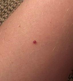
© Midasblenny, CC BY-SA 4.0, a través de Wikimedia Commons
Máculas de 1-3 mm de diámetro que pueden convertirse en pápulas más grandes con el tiempo.
Color rojo cereza brillante típico, pero puede parecer azul o púrpura.
They are non-blanching.
Continúa leyendo abajo
Presentación
Volver al contenidoUsualmente ocurren en el tronco y las extremidades superiores.
Se pueden encontrar en cualquier parte de la piel, excepto en las membranas mucosas. Se ha informado del cuero cabelludo.1
Las lesiones pueden ser generalizadas, especialmente en los ancianos.
Por lo general, son asintomáticos.
Diagnóstico diferencial
Volver al contenidoEl diagnóstico suele ser claro clínicamente. El diagnóstico diferencial puede incluir:
Angiokeratoma.
Lagos venosos (angiomas azules, más a menudo en los labios).
Gestión de manchas de Campbell de Morgan
Volver al contenidoTranquilizar - estas lesiones generalmente no requieren tratamiento.
Muy ocasionalmente, puede ser necesario extirparlas si las lesiones se enganchan o por razones estéticas.
Si se desea la eliminación, las opciones de tratamiento incluyen curetaje, láser de colorante pulsado, electrocauterización y escisión.
Se ha encontrado que la escleroterapia también es efectiva.3
Cuándo derivar
Volver al contenidoCuando hay incertidumbre diagnóstica.
Cuando se requiere asistencia para la eliminación.
Pronóstico
Volver al contenidoLas manchas de Campbell de Morgan son lesiones benignas.
Los problemas solo surgen cuando las lesiones son frecuentemente traumatizadas, continúan agrandándose o son de preocupación estética para un paciente.
Exclusive updates for healthcare professionals
Stay informed with the latest clinical updates, professional insights, and evidence-based guidance. The Patient Pro newsletter curates essential content for healthcare professionals—delivered straight to your inbox.
By subscribing you accept our Política de Privacidad. Puedes darte de baja en cualquier momento. Nunca vendemos tus datos.
Lecturas adicionales y referencias
- Angioma Senil; DermIS (Sistema de Información de Dermatología)
- Higgins JC, Maher MH, Douglas MS; Diagnóstico de Tumores Cutáneos Benignos Comunes. Am Fam Physician. 1 de octubre de 2015;92(7):601-7.
- Angioma (adquirido) - incluyendo angioma cereza / manchas de Campbell de Morgan; Sociedad de Dermatología de Atención Primaria (PCDS)
- Kim JH, Park HY, Ahn SK; Angiomas de Cereza en el Cuero Cabelludo. Caso Rep Dermatol. 2009 Nov 11;1(1):82-86.
- Angiomas; DermNet NZ
- Jairath V, Dayal S, Jain VK, et al; ¿Es útil la escleroterapia para los angiomas de cereza? Dermatol Surg. 2014 Sep;40(9):1022-7. doi: 10.1097/01.DSS.0000452631.83962.58.
Continúa leyendo abajo
About the authorView full bio

Dra. Rachel Hudson, MRCGP
General Practitioner and Medical Author
MBChB, MRCGP (2008), BSc (Medical Science), DFSRH, DRCOG, DCH
Dr Rachel Hudson, is an NHS GP working in the North West of England.
About the reviewerView full bio

Dra. Toni Hazell, MRCGP
MBBS, BSc, MRCGP, DFSRH, Dip GU med, DRCOG, DCH (London, UK, 2000)
Dr. Toni Hazell qualified from St. Mary’s Hospital Medical School and did her VTS at Northwick Park Hospital.
Historial del artículo
La información en esta página está escrita y revisada por pares por clínicos calificados.
Próxima revisión: 30 de octubre de 2027
31 Oct 2024 | Última versión

Pregunta, comparte, conecta.
Navega por discusiones, haz preguntas y comparte experiencias en cientos de temas de salud.

¿Te sientes mal?
Evalúa tus síntomas en línea de forma gratuita